
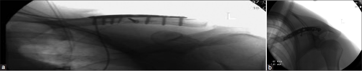
Figure 3: (a) Fluoroscopic image angled 30° superiorly (cephalic). (b) Fluoroscopic image angled 60° inferiorly (caudal)
|
|
Close |
|
Figure 3: (a) Fluoroscopic image angled 30° superiorly (cephalic). (b) Fluoroscopic image angled 60° inferiorly (caudal)
|
|