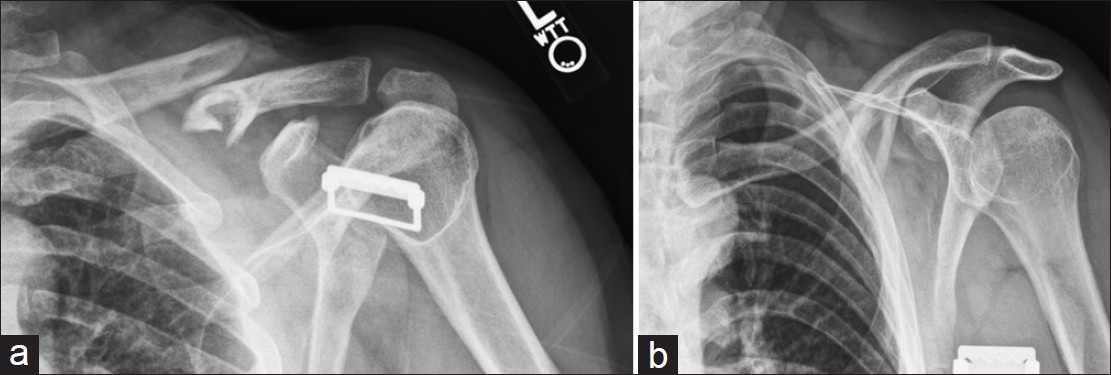
Figure 5: (a and b) Orthogonal views of comminuted, segmental clavicle fracture. The AP view illustrates approximately 100% displacement with an irregular complex appearance of the medial aspect of the lateral fracture fragment, not clearly depicting this area. The 60° caudally angled view identifies a segmental fragment that is angled more in the sagittal plane, plainly seen as the round tubular structure of the midshaft of the clavicle on the traditional clavicle AP view. This segmental fragment could have been easily missed without the former view