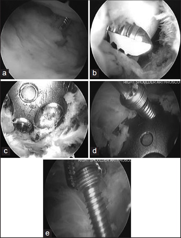
Figure 1: Right shoulder arthroscopic images obtained from a posterior portal using a 30° arthroscope and performed in the lateral decubitis position, in a patient undergoing screw removal. Diagnostic glenohumeral arthroscopy reveals the prominent screw (a) and associated glenoid erosion (b). Through subacromial bursoscopy, the plate is exposed after excision of scar tissue with a combination of a radiofrequency device, a shaver and a labral elevator (c). The screw to be removed is identified (d) and grasped with a needle holder as it is withdrawn (e)