
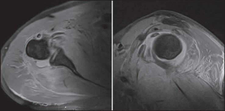
Figure 1: Axial and sagittal fat suppressed T2 weighted MR images displaying joint effusion and high signal change within the muscle bellies of subscapularis and teres minor
|
|
Close |
|
Figure 1: Axial and sagittal fat suppressed T2 weighted MR images displaying joint effusion and high signal change within the muscle bellies of subscapularis and teres minor
|
|