
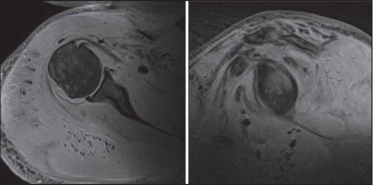
Figure 2: Axial and sagittal fat suppressed T2 weighted MR images displaying further high signal change within the muscle bellies in addition to low signal loculations of gas within the soft tissues
|
|
Close |
|
Figure 2: Axial and sagittal fat suppressed T2 weighted MR images displaying further high signal change within the muscle bellies in addition to low signal loculations of gas within the soft tissues
|
|