
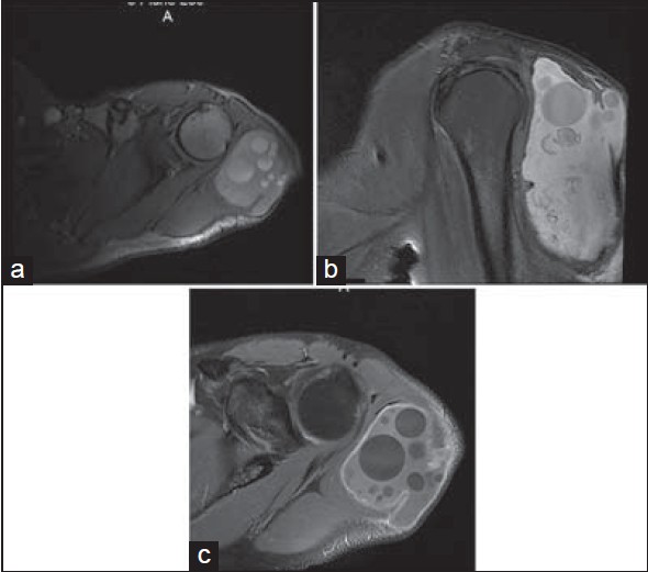
Figure 2: MRI aspects of T1 weighted axial image (a) and sagittal image (b) with fat suppression of the cyst, also T2-weighted coronal image shows the daughter cysts well (c)
|
|
Close |
|
Figure 2: MRI aspects of T1 weighted axial image (a) and sagittal image (b) with fat suppression of the cyst, also T2-weighted coronal image shows the daughter cysts well (c)
|
|