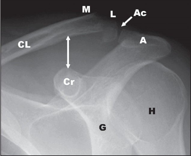
Figure 1: A 15° cephalad tilt radiograph of the glenohumeral joint shows a noncomminuted lateral clavicle fracture. Increased coracoclavicular distance (arrows) suggests disruption of the coracoclavicular ligaments. (Ac: acromioclavicular joint, A: acromion, CL: clavicle, M: medial fragment, L: lateral clavicle fragment, Cr: coracoid, G: glenoid, H: humeral head)