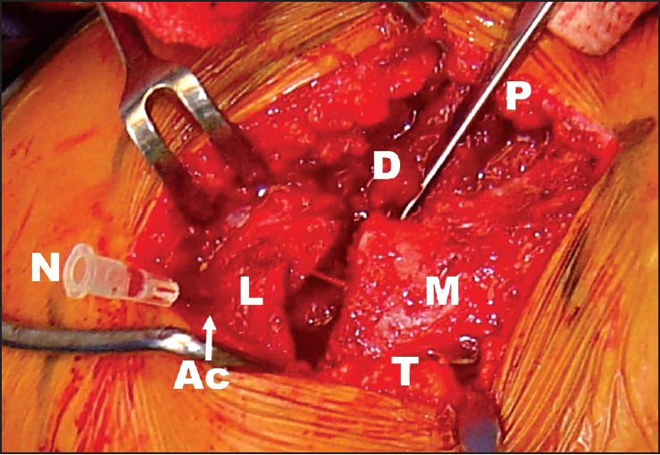
Figure 2: Intraoperative demonstration of the pathoanatomy of lateral clavicle fractures. A needle (N) marks the position of the acromioclavicular joint (Ac). The torn deltoid (D) and trapezius (T) attachments and the lateral (L) and medial (M) clavicular fragments are seen. A probe (P) is used to palpate the coracoid process