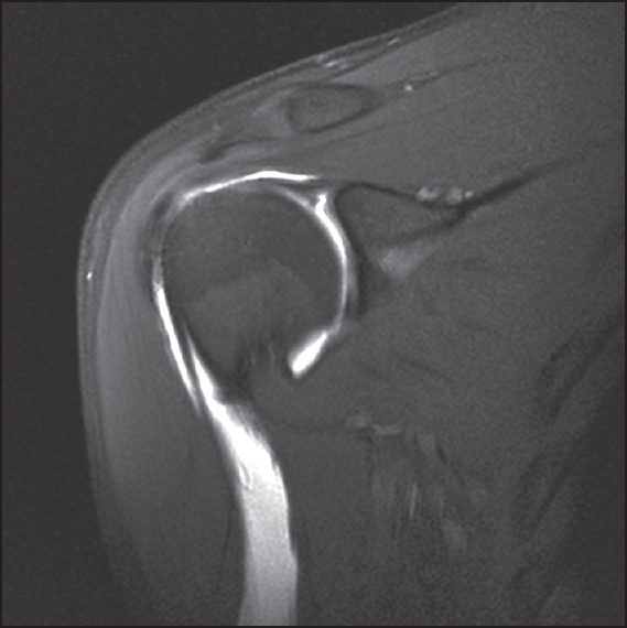
Figure 3: T1-weighted, fat-suppressed, coronal oblique magnetic resonance imaging arthrography images demonstrating a patient with a recurrent type II SLAP lesions following type II SLAP repair. Note the high signal (gadolinium) present between the superior labrum and glenoid present postoperatively