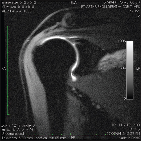
Figure 4: T1-weighted, fat-suppressed, coronal oblique magnetic resonance imaging arthrography images demonstrating a patient with an intact superior labrum following type II SLAP repair. Note the absence of gadolinium between the superior labrum and glenoid postoperatively