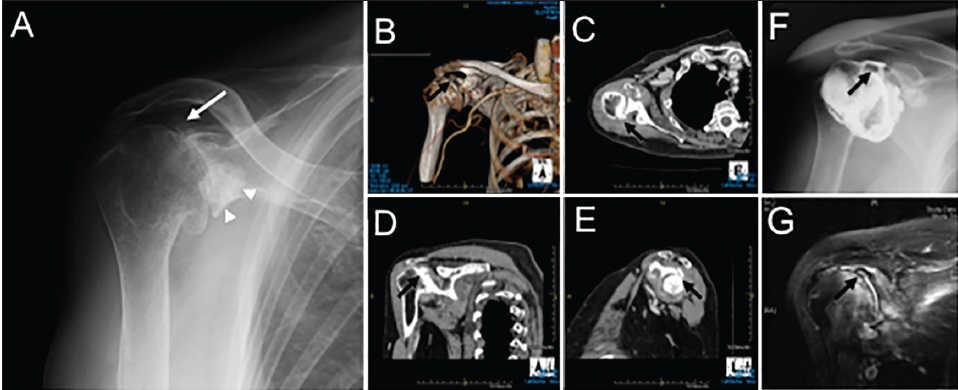
Figure 1: Antero-posterior radiographs of right shoulder (a). Three-dimension (b), Axial (c), Oblique coronal (d), and oblique sagittal (e) Views of computed tomography of the right shoulder. Antero-posterior view of arthrography (f) and oblique coronal view of enhanced magnetic resonance imaging (g) of the right shoulder. Arrows indicate a small bone fragment at the superior-posterior glenoid rim. Arrowheads showing large bone fragments under the bilateral coracoid processes