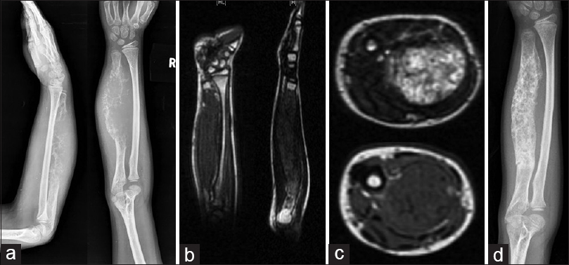
Figure 1: (a) Plain radiograph of forearm showing aggressive lesion of ulna with cortical destruction and soft tissue component. (b and c) Magnetic resonance imaging showing craniocaudal extent of the disease in ulna and the relation of the tumor with the neurovascular bundle. (d) Plain radiograph of forearm after neoadjuvant chemotherapy showing a good response