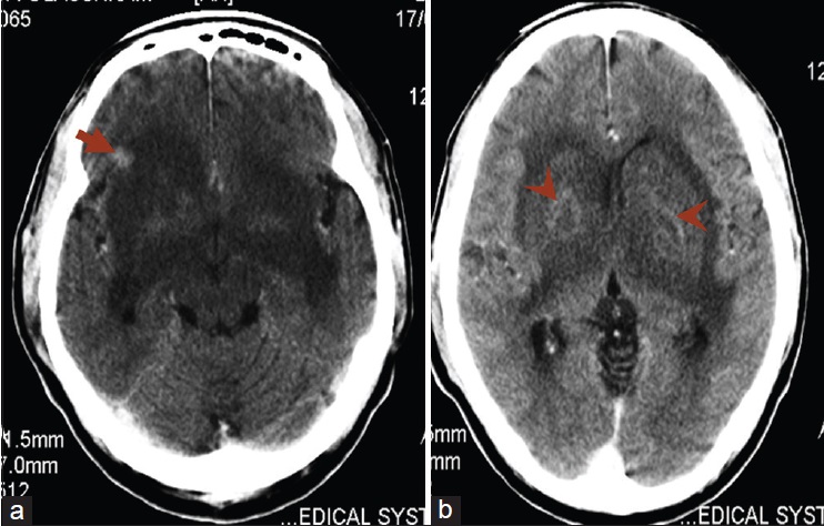
Figure 1: Axial contrast enhanced computed tomography of the brain: (a) An area of extensive hypodensity in the region of the basal ganglia bilaterally with compression of the third ventricle and a focus of enhancing hypodensity in right frontal lobe (arrow), (b) anterior extension of the hypodensity with resultant compression of the anterior horns of both lateral ventricle with ring enhancement within the hypodensity in the basal ganglia (arrowheads)