|
 
 |
| ORIGINAL ARTICLE |
|
| Year : 2014 | Volume
: 2
| Issue : 1 | Page : 11-16 |
|
Assessment of skeletal maturity using the permanent mandibular canine calcification stages
Sandeep Goyal1, Sonia Goyal2, Neeraj Gugnani3
1 Dental Department, King Faisal Hospital, Affiliate of the National University of Rwanda, Kigali, Rwanda
2 Consultant Stomatologist and Maxillofacial Surgeon, Polyclinique La Medicale, Kigali, Rwanda
3 Department of Pedodontics, DAV C Dental College, Yamuna Nagar, Haryana, India
| Date of Web Publication | 29-Jan-2014 |
Correspondence Address:
Sandeep Goyal
Senior Consultant Orthodontist, Dental Department, King Faisal Hospital, affiliate of National University of Rwanda
Rwanda
 Source of Support: None, Conflict of Interest: None  | Check |
DOI: 10.4103/2321-3825.125916

Aim: The aim of this study is to assess, (1) the relationship between cervical vertebrae maturation and mandibular canine calcification stages; and (2) whether the mandibular canine calcification stages can be used as indicators to determine skeletal maturity. Materials and Methods: A descriptive, retrospective, and cross-sectional study was designed. A null hypothesis was proposed that there was no relationship between cervical vertebrae maturation and the mandibular canine calcification stages. Pre-treatment orthopantomograms (OPGs) and lateral cephalograms of 99 males and 110 females of Rwanda ethnicity were selected. The cervical vertebrae maturation index (CVMI) proposed by Hassel and Farman was used to evaluate the skeletal maturation level, and the mandibular canine calcification stages were assessed with the Demirjian Index (DI). Results: A significant association was found between the CVMI and DI stages, as evaluated by the Pearson contingency coefficient values (0.599 for males and 0.719 for females). Canine stage F in males and canine stage E in females could represent the CVMI 2 stage and indicate the onset of a period of accelerating growth. Conclusions: The mandibular canine calcification stages might be clinically used as maturity indicators of the pubertal growth period, but only during the onset and accelerating phases. Keywords: Cervical vertebrae, Demirijan index, mandibular canine, orthodontics, skeletal maturity
How to cite this article:
Goyal S, Goyal S, Gugnani N. Assessment of skeletal maturity using the permanent mandibular canine calcification stages. J Orthod Res 2014;2:11-6 |
How to cite this URL:
Goyal S, Goyal S, Gugnani N. Assessment of skeletal maturity using the permanent mandibular canine calcification stages. J Orthod Res [serial online] 2014 [cited 2018 Mar 20];2:11-6. Available from: http://www.jorthodr.org/text.asp?2014/2/1/11/125916 |
| Introduction | |  |
Knowledge of the status of the pubertal growth spurt of an individual is important for a clinician, as it influences diagnosis, treatment planning, and the eventual outcome of the orthodontic treatment. [1],[2] The skeletal maturity can be assessed by inspection of the ossification-related changes in the hand-wrist and the cervical vertebrae regions assessed on the radiographs. [3] The hand-wrist radiograph has been commonly used for skeletal maturity assessment. [1] The cervical vertebrae maturation indicators on the lateral cephalograms have also been found to be reliable skeletal maturation predictors. [3]
Studies have proposed the assessment of dental calcification stages to determine the level of skeletal maturation. It was concluded that a strong relation exists between dental maturity and skeletal maturity to evaluate the skeletal growth status. [4],[5] Relationships between the calcification stages of various mandibular teeth and skeletal maturity have been reported, with variable significance. [1] Mandibular canine calcification has been found to be a reliable indicator in some studies, [6],[7],[8] while others have refuted the claim. [9],[10],[11]
The objective of this study was to investigate the relationship between the stages of cervical vertebrae maturation and permanent mandibular canine calcification. A null hypothesis was proposed that there was no significant relationship between cervical vertebrae maturation and the mandibular canine calcification stages.
| Materials and Methods | | |
It was a descriptive, retrospective, and cross-sectional research project. Good quality, pretreatment OPGs and lateral cephalograms of 209 subjects (99 males and 110 females) of Rwanda ethnicity were selected from the records of orthodontic patients.
Selection criteria : The inclusion criteria were as follows:
- Chronological age ranging from seven to nineteen years
- Normal overall growth and development
- Absence of abnormal dental conditions, such as, impaction, transposition, and congenitally missing teeth
- Absence of a previous history of trauma or disease to the face and neck
- No gross skeletal deformities, for example, clefts, hemiatrophy, hypertrophy, and so on, and no congenital deformity
- Absence of previous orthodontic treatment
- No permanent teeth extracted
All radiographic assessments were performed by a single examiner on a back-illuminated radiographic view box in a darkened room.
Evaluation of cervical vertebrae maturity (CVMI) on a lateral cephalogram: The CVMI was evaluated by using the method proposed by Hassel and Farman [3] [Table 1].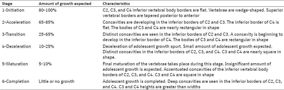 | Table 1: Cervical vertebrae maturation indicators (CVMI, Hassel and Farman, 1995)
Click here to view |
Evaluation of dental maturity (DI) on a panoramic radiograph: The development stages of mandibular left canine were assessed according to Demirjian et al.[12] (Demirjian Index, DI) [Table 2]. If the left side tooth was not clear, then the right side tooth was evaluated.
Randomly selected records of 15 patients were re-evaluated after two weeks of the first evaluation, to test the reproducibility of the assessments of DI and CVMI, and the data was evaluated in terms of the weighted kappa statistics for DI and CVMI. The kappa statistics for intraobserver agreement were 0.82 for DI assessments and 0.89 for CVMI assessments, showing acceptable intraobserver agreement during assessments.
Statistical Analysis
The statistical analyses were performed with SPSS 13.0, SPSS Inc, Chicago, III., and Epi Info 3.4.3 (CDC, Illinois). Descriptive statistics were applied for both genders to determine the percentage distribution of subjects, the means and standard deviations of the chronological ages for the stages of CVMI, and DI stages of mandibular canines. Cross-tabulation was done to find the distribution of the DI stages among the CVMI stages, stratified by the gender. Mann Whitney / Wilcoxon Two-sample test (Kruskal Wallis test for two groups), the Pearson chi-square test values, and Pearson contingency coefficient were also estimated to determine the relationship between DI and CVMI. A p-value of < 0.01 was considered as statistically significant.
| Results | | |
The study involved the records of 47.4% (n = 99) males and 52.6% (n = 110) females. The age range of the study sample was from seven years to eighteen years seven months. The mean age of the males was 13.27 years (SD = 2.1 years), of females was 12.89 years (SD = 2.26 years), and that of the total sample as 13.08 years (SD = 2.18 years), [Table 3].
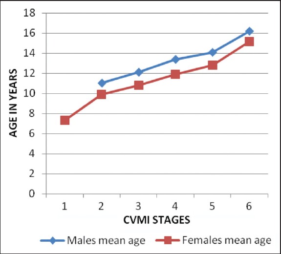
[Table 4] shows the percentage distribution and mean age at different CVMI stages. Each stage appeared earlier in females than in males, consistently [Figure 1]. Mann Whitney / Wilcoxon Two-sample test (Kruskal Wallis test for two groups) showed highly significant differences among various CVMI stages in both the genders (Females, Chi square χ2 (5) = 81.57, P < 0.001; and Males, χ2 (4) = 59.08, P < 0.001). 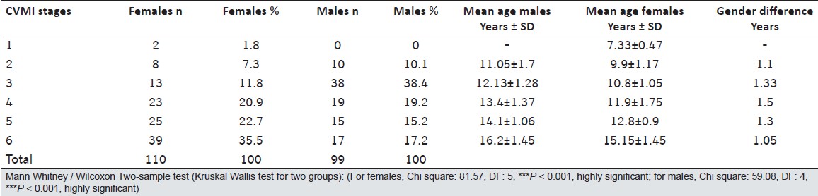 | Table 4: Percentage distribution and the mean chronological ages of all the subjects grouped by various CVMI stages
Click here to view |
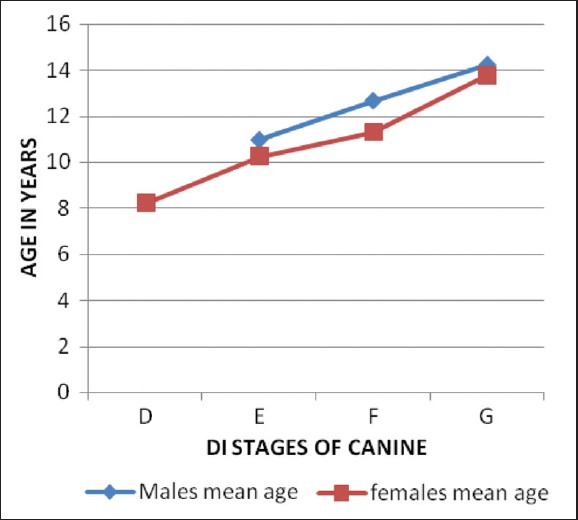
[Table 5] shows the mean age at different DI stages of mandibular canines. Females achieved subsequent maturation stage of mandibular canines earlier [Figure 2]. Mann Whitney / Wilcoxon Two-sample test (Kruskal Wallis test for two groups) showed highly significant differences among various DI stages in both the genders (Females, χ2 (3) = 50.74, P < 0.001; Males, χ2 (2) = 40.67, P < 0.001).
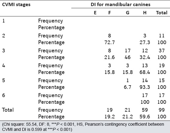
[Table 6] shows the association and distribution between the DI stages of mandibular canines and CVMI stages in males. The χ2 (8) was highly significant at 55.54 (P < 0.001). The Pearson contingency coefficient was 0.599, showing a significant association between DI and CVMI. In males, the DI Stage F showed the highest percentage distribution (73%) at stage 2 of CVMI (pre-peak of pubertal growth spurt) and DI stage G showed a large percentage distribution in stage 3 of CVMI (peak of pubertal growth spurt). The DI stage H was associated with stages 4, 5, and 6 of CVMI (end of pubertal growth spurt).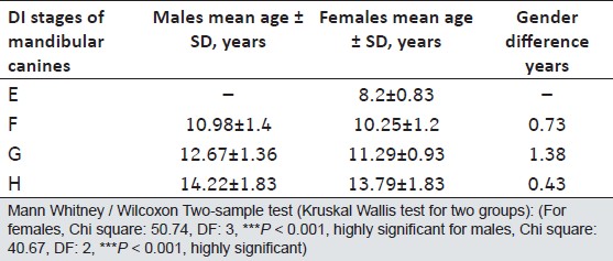 | Table 5: Mean age of different DI stages of mandibular canines in the genders and the gender difference
Click here to view |
 | Table 6: Contingency table showing association and distribution between DI stages of mandibular canines and CVMI stages for males
Click here to view |
[Table 7] shows the association and distribution between the DI stages of mandibular canines and CVMI stages in females. The χ2 (15) was highly significant at 117.48 (p < 0.001). The Pearson contingency coefficient was 0.719 showing a highly significant association between DI and CVMI. The statistical values for female subjects were higher than for males, showing a stronger association in females. 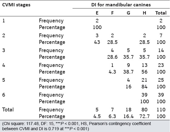 | Table 7: Contingency table showing association and distribution between DI stages of mandibular canines and CVMI stages for females
Click here to view |
Thus the null hypothesis was rejected and a significant relationship was found to exist between the CVMI stages and mandibular canine DI stages in both the genders. In females, the DI stage E corresponded to CVMI stages 1 and 2; DI stage G with CVMI stage 3, and DI stage H with CVMI stages 4, 5, and 6. However, there was a scattered distribution of canine DI stages in females in the CVMI 3 stage, while it was better in males. A comparison of [Table 6] and [Table 7] also shows that the DI in male subjects was more advanced than in female subjects with respect to the CVMI stages.
| Discussion | | |
OPGs were chosen for dental maturity assessment, as they are routinely available in orthodontic clinics, and the mandibular region is clearly visible. The Demirjian et al.[12] method was chosen for dental maturation assessment in the present study, because it is based on shape criteria and proportion of root length, using the relative value to crown the height rather than the absolute length. Therefore, the foreshortened or elongated projections of developing teeth will not affect their reliability of assessment. [1]
The skeletal age assessment has been assessed on the lateral cephalograms, using the CVMI method described by Hassel and Farman. [3] It is quick and relatively easy to perform. It omits the hand-wrist x-ray, thus avoiding excessive radiation exposure. Studies have found that CVMI is a reliable method for skeletal maturity assessment, equivalent to the hand-wrist x-rays, and shows greater reproducibility between the observers. [3],[13],[14]
According to Hassel and Farman, [3] CVMI 2 stage indicates the onset of accelerating growth velocity, CVMI 3 shows a period of rapid growth velocity, while CVMI 4 denotes the period of decelerating growth. The mean age for each CVMI skeletal maturity level presented in [Table 4] indicated that females mature earlier than males by an average of 1.5 years. This finding is in corroboration with other studies. [1],[15],[16],[17],[18] Rwandese children and adolescents seem to be a little more advanced in skeletal maturation, as compared to other population groups. [19]
Dental and Skeletal Maturity
The females were more advanced than the males in both dental and skeletal maturation, which was in accordance with the previous reports. [7],[8],[9],[13] However, the maturation patterns of tooth development in relation to CVMI skeletal maturity, was more advanced in males as compared to females [Table 6] and [Table 7]. At the same skeletal maturity stage, males had a higher distribution of the later dental developmental stages. This result was similar to the findings of previous studies. [1],[8],[20],[21],[22] It is therefore suggested that tooth mineralization relative to the stages of skeletal maturation should be considered individually for both genders.
In this study, girls were found to mature earlier than boys, which was in agreement with the previous studies. [1],[8],[21],[22],[23] Dental maturation also occurred earlier in girls than in boys, by almost a year, and completed earlier in girls. This suggested that a sexual dimorphism existed in mandibular canine tooth formation. However, Chertkow and Fatti [24] reported no significant sex differences, while other studies reported an advanced trend in boys compared to girls. [1],[8],[20] Conversely, Basaran et al. stated that boys showed late dental development. [13]
The Pearson correlation coefficients between skeletal maturity and canine calcification stages in our study were 0.719 in females and 0.599 in males (p < 0.001), which confirmed the previous findings. [1],[6],[7],[8],[20] It suggested that a moderate association existed between the skeletal and dental maturation stages.
In our study, DI stage F was coinciding well with CVMI stage 2 in males, but there was a scattered distribution of canine calcification stages in females in the CVMI 2 stage. Divyashree et al.[9] found Stage F to be coinciding well with the pre-peak of skeletal development in the Indian sample, while Coutinho et al. found that in children from USA, the canine stage G was more indicative of the pre-peak of the growth spurt. [7] Chertkow [6] and Fatti and Chertkow [20] had previously reported mandibular canine stage G coinciding with the early appearance of the sesamoid (which was equivalent to CVMI 2) [3] in boys as well as in girls, while Krailassikari [1] and So [25] found no uniformity in canine development in the same skeletal maturity stage.
In our study, in CVMI 3 stage, the majority of the males was at DI stage G, while there was a scattered distribution of canine stages in females. At CVMI stages 4 and 5, in both genders, most of the canines were in stage G and H, that is, toward root completion, while 100% canines were in stage H at the CVMI 6 stage. It showed that canine stages could be considered as indicative of growth status in the beginning stages (stages E and F) and has more reliable significance in males than females. This was in accordance with the findings of Divyashree et al.[9] in Indian subjects and Lu [10] in Chinese subjects for the males.
It signifies that the interpretation of the relationship between mandibular canine calcification stages with the later stages of skeletal maturity is not meaningful. Mandibular canine calcification stages can only be used reliably in their earlier stages for predicting skeletal maturation levels. We have observed that there is no uniform distribution of the sample in the different stages of DI, which can be due to the small sample size and a larger age range of the study subjects. Future studies are recommended with a larger sample size and reduced age range. Also, a uniform number of study subjects must be selected in different age categories.
Clinical Implications
Knowledge of active growth is important for clinical decisions, especially for dentofacial orthopedic, surgical, and prosthetic treatment planning. Studies have reported that there is a greater skeletal response with myofunctional appliances if treatment is given during the peak height velocity period than during the pre-peak period. [1] Thus for more orthopedic effects, the treatment must be started during the CVMI 2-3 stages. Treatment given after these stages may result in more dental than skeletal effects. [1]
The relationship between the mandibular canine calcification stages and the skeletal maturity indicators can allow the clinician to more easily identify stages of the pubertal growth period. We found that the canine stage F in males and canine stage E in females may represent the CVMI 2 stage and can serve as a simple tool for evaluating the onset of the accelerating growth period. It can be easily incorporated into clinical practice by using the intra-oral periapical (IOPA) view for initial growth assessment of an individual.
| Conclusion | | |
A significant relation exists between the mandibular canine calcification and skeletal development, which is stronger in females than males. Canine stage F in males and canine stage E in females may represent the CVMI 2 stage and indicate the onset of a period of accelerating growth. The findings of this study indicate that the mandibular calcification stages may be clinically used as a maturity indicator of the pubertal growth period, but only during the onset and accelerating phases. However, further study is recommended in a larger sample to corroborate the findings and to evaluate the mandibular maturation of the other teeth in relation to skeletal maturity.
| References | | |
| 1. | Krailassiri S, Anuwongnukroh N, Dechkunakorn S. Relationships between dental calcification stages and skeletal maturity indicators in Thai individuals. Angle Orthod 2002;72:155-65. 
[PUBMED] |
| 2. | Krogman WM. Biological timing and the dento-facial complex. ASDC J Dent Child 1968;35:175-85, 328-41, 377-81.
[PUBMED] |
| 3. | Hassel B, Farman AG. Skeletal maturation evaluation using cervical vertebrae. Am J Orthod Dentofac Orthop 1995;107: 58-66.
|
| 4. | Morris JM, Park JH. Correlation of dental maturity with skeletal maturity from radiographic assessment: A review. J Clin Pediatr Dent 2012;36:309-14.
[PUBMED] |
| 5. | Mittal S, Singla A, Virdi M, Sharma R, Mittal B. Co-relation between determination of skeletal maturation using cervical vertebrae and dental calcification stages. Internet J Forensic Sci 2011;4:1-8
|
| 6. | Chertkow S, Fatti P. Relationship between tooth mineralization and early radiographic evidence of ulnar sesamoid. Angle Orthod 1979;49:282-8.
[PUBMED] |
| 7. | Coutinho S, Buschang PH, Miranda F. Relationship between mandibular canine calcification stages and skeletal maturity. Am J Orthod 1993;104:262-8.
|
| 8. | Uysal T, Sari Z, Ramoglu SI, Basciftci FA. Relationships between dental and skeletal maturity in Turkish subjects. Angle Orthod 2004;74:657-63.
[PUBMED] |
| 9. | Divyashree R, Dinesh MR, Amarnath BC. Reliability of permanent mandibular canine calcification as an indicator of skeletal maturity in Karnataka population. World J Dent 2010;1:5-9.
|
| 10. | Lu Y. Relationships between mandibular canine calcification stages and skeletal maturity. Zhonghua Kou Qiang Yi Xue Za Zhi 1999;34:40-2.
|
| 11. | Mappes MS, Harris EF, Behrents RG. Regional differences in tooth and bone development. Am J Orthod Dentofacial Orthop 1992;101:145-51.
|
| 12. | Demirjian A, Goldstein H, Tanner JM. A new system of dental age assessment. Hum Biol 1973;45:211-27.
[PUBMED] |
| 13. | Basaran G, Ozer T, Hamamci N. Cervical vertebral and dental maturity in Turkish subjects. Am J Orthod Dentofacial Orthop 2007;131:447.e13-20.
|
| 14. | Baccetti T, Franchi L, Cameron CG, McNamara JA. Treatment timing for rapid maxillary expansion. Angle Orthod 2001;71:343-50.
|
| 15. | Fishman LS. Maturational patterns and prediction during adolescence. Angle Orthod 1987;57:178-93.
[PUBMED] |
| 16. | Bjork A, Helm S. Prediction of the age of maximum pubertal growth in body height. Angle Orthod 1972;37:134-43.
|
| 17. | Grave KC, Brown T. Skeletal ossification and the adolescent growth spurt. Am J Orthod 1976;69:611-9.
[PUBMED] |
| 18. | Hagg U, Taranger J. Maturation indicators and the pubertal growth spurt. Am J Orthod 1982;82:299-309.
|
| 19. | Soegiharto BM, Moles DR, Cunningham SJ. Discriminatory ability of the skeletal maturation index and the cervical vertebrae maturation index in detecting peak pubertal growth in Indonesian and white subjects with receiver operating characteristics analysis. Am J Orthod Dentofacial Orthop 2008;134:227-37.
[PUBMED] |
| 20. | Chertkow S. Tooth mineralization as an indicator of the pubertal growth spurt. Am J Orthod 1980;77:79-91.
[PUBMED] |
| 21. | Demirjian A, Levesque GY. Sexual differences in dental development and prediction of emergence. J Dent Res 1980;59:1110-22.
[PUBMED] |
| 22. | Chapman SM. Ossification of the adductor sesamoid and adolescent growth spurt. Angle Orthod 1972;42:236-44.
[PUBMED] |
| 23. | Fishman LS. Radiographic evaluation of skeletal maturation. Angle Orthod 1982;52:88-111.
[PUBMED] |
| 24. | Chertkow S, Fatti P. The relationship between tooth mineralization and early evidence of the ulnar sesamoid. Angle Orthod 1979;49:282-8.
[PUBMED] |
| 25. | So LL. Skeletal maturation of the hand and wrist and its correlation with dental development. Aust Orthod J. 1997;15:1-9.
|
[Figure 1], [Figure 2]
[Table 1], [Table 2], [Table 3], [Table 4], [Table 5], [Table 6], [Table 7]
|


 Search Pubmed for