|
 
 |
| ORIGINAL ARTICLE |
|
| Year : 2014 | Volume
: 2
| Issue : 3 | Page : 125-128 |
|
Investigation of the peg-shaped maxillary lateral incisors in a Turkish orthodontic subpopulation
M Karatas1, MS Akdag2, Mevlut Celikoglu3
1 Department of Restorative Dentistry, Faculty of Dentistry, Recep Tayyip Erdogan University, Rize, Turkey
2 Department of Operative Dentistry, Faculty of Dentistry, Akdeniz University, Trabzon, Turkey
3 Department of Orthodontics, Faculty of Dentistry, Akdeniz University, Trabzon, Turkey
| Date of Web Publication | 12-Sep-2014 |
Correspondence Address:
Mevlut Celikoglu
Department of Orthodontics, Faculty of Dentistry, Akdeniz University, Trabzon
Turkey
 Source of Support: None, Conflict of Interest: None  | Check |
DOI: 10.4103/2321-3825.140675

Aim: The following study aims to investigate the prevalence and distribution of the peg-shaped maxillary lateral incisors in a Turkish orthodontic subpopulation. Materials and Methods: A study sample consisted of 2925 patients of which 1746 were females and 1179 were males with aged range from 10 to 18 years; (mean age, 13.40 ± 1.9 years) were retrospectively examined using panoramic radiographs, dental cast and pre-treatment records for the presence of the peg-shaped maxillary lateral incisors. Pearson's Chi-square and Fisher exact tests were used for statistical comparisons. Results: Peg-shaped maxillary lateral incisors were found in 2.15% of the patients (63/2925; 41 females and 22 males) with no statistically significant difference between the genders. However, females had more peg-shaped maxillary left lateral incisors compared with the males (P = 0.007). It was almost equally distributed as bilateral (1.09%, 32/2925) or unilateral (1.06%, 31/2925). Conclusion: It was found to be 2.15% (63 out of 2925 patients) with no gender difference except for the peg-shaped maxillary left lateral incisors. Clinicians should be aware of this anomaly prior to treatment for possible complications. Keywords: Peg-shape, peg shaped maxillary laterals, prevalence
How to cite this article:
Karatas M, Akdag M S, Celikoglu M. Investigation of the peg-shaped maxillary lateral incisors in a Turkish orthodontic subpopulation. J Orthod Res 2014;2:125-8 |
How to cite this URL:
Karatas M, Akdag M S, Celikoglu M. Investigation of the peg-shaped maxillary lateral incisors in a Turkish orthodontic subpopulation. J Orthod Res [serial online] 2014 [cited 2018 Feb 10];2:125-8. Available from: http://www.jorthodr.org/text.asp?2014/2/3/125/140675 |
| Introduction | |  |
Dental anomalies in tooth number, size, morphology, or eruptive pattern of the teeth may be congenital, developmental, or acquired. Some of these abnormalities may also occur by a combination of both hereditary and environmental factors. [1],[2] For over 40 years, the literature has suggested that the occurrence of peg-shaped lateral incisors was associated with the same genetic mechanism that causes agenesis of the maxillary lateral incisors. [3],[4] Peg-shaped lateral incisor teeth can be unilateral or bilateral in same patients. The identification of these teeth indicates the diagnosis. [2]
Jukic et al. [5] believe that the incidence of dental anomalies can provide important information for phylogenic and genetic studies and help to understand variations within and between populations. The prevalence of peg-shaped maxillary lateral incisors is not uniform and varies according to the population. Prevalence rates of peg-shaped lateral incisors have been reported to a range from 0.6% [6] to 9.9% [7] in different populations.
Although the prevalence of different dental anomalies in different populations has been reported in several studies, there were few studies involving peg-shaped permanent lateral incisors in Turkish population. Altug-Atac and Erdem [8] evaluated the prevalence of dental anomalies in 3043 orthodontic patients and reported that 48 patients had peg-shaped maxillary and mandibular lateral incisors. In a study by Kazanci et al. [9] investigated the prevalence of different developmental dental anomalies and reported that prevalence rates of peg-shaped maxillary lateral incisors were 2.12%.
Anomalies in size may lead to disturbances in maxillary and mandibular arch length and occlusion [7] and may also cause aesthetic and psychological problems. Therefore, peg-shaped tooth is a major concern among dentists. The aim of this study was to determine the prevalence and distribution of peg-shaped maxillary lateral incisors in a Turkish orthodontic subpopulation and compare the findings with different populations.
| Materials and Methods | | |
The patients included in the present study were not exposed to any additional radiation particularly for this retrospective study. Thus, further ethics approval was not required for the present retrospective archieve study. In addition, as a usual protocol, all the patients (or parents) signed an informed consent agreeing to the use of the patients' data for scientific studies.
Records of patients with medical problems, history of extraction of any permanent tooth except third molars, cleft lip and palate, trauma were excluded from study (25 patients). Finally, the data of 2925 orthodontic patients (1746 females and 1179 males), who attended to the Karadeniz Technical and Recep Tayyip Erdogan Universities were examined for the presence of peg-shaped maxillary lateral incisors. The mean age of the patients ranged from 10 to 18 years with a mean age of 13.40 ± 1.9 years.
Peg-shaped maxillary lateral incisors presenting variations tooth size and number were evaluated as conical crown-size reduction or the mesiodistal width of an incisor tooth being shorter than the cervical width of the tooth crown according to criteria defined by Langlais et al. [10] Mesiodistal diameters of the maxillary lateral incisors were measured using a digital caliper (Mitutoyo, Tokyo, Japan). Then data obtained from dental cast were recorded as unilateral (left or right) or bilateral, according to gender and number of patients with peg-shaped maxillary lateral incisors.
Statistical Analyses
The Statistical Package for Social Sciences program to analyze data (SPSS, version 16.0, Chicago, IL, USA) was used. Student's t-test was used to compare the chronological ages between genders. The patterns of peg-shaped maxillary lateral incisors were tested using the Pearson's Chi-square and Fisher exact tests at the 5% significance level for a possible gender difference.
| Results | | |
The number of females in our study (59.7%) was more than the males (40.3%). There was no statistically significant difference between the females (13.44 ± 1.96 years) and males (13.33 ± 1.86 years) in terms of chronological ages [Table 1].
Peg-shaped maxillary lateral incisors were found in 2.15% of the examined patients (63/2925; 41 females and 22 males). There was no statistically difference between the genders, while prevalence rates for female patients (2.35%) were higher than for male patients (1.87%). Females had more peg-shaped left and right maxillary lateral incisor (0.97% and 0.46%, respectively), while males had more peg-shaped bilateral maxillary lateral incisors (1.36%). There was a statistically significant difference between females and males for the peg-shaped maxillary left lateral incisors (P = 0.007). Peg-shaped maxillary lateral incisors were almost equally distributed as bilateral (1.09%, 32) or unilateral (1.06%, 31). Among the unilateral peg-shaped laterals, left side ones (0.65%) were more common than the right-side ones (0.41%) [Table 2].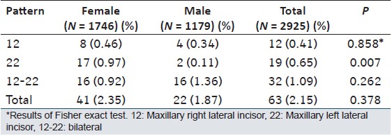 | Table 2: Prevalence and distribution of the peg shaped maxillary lateral incisors
Click here to view |
| Discussion | | |
Peg-shaped maxillary lateral incisors are a major concern of general dentists and dental specialist since they cause esthetic, orthodontic and periodontal problems for affected patients. The prevalence of peg-shaped lateral incisors might be helpful in discovering the genetic and environmental causes for peg-shaped lateral formation and also establishing a basic awareness among dentists. [11]
In our country, few studies reporting the prevalence of dental anomalies evaluated the peg-shaped maxillary lateral incisors in orthodontic patients and the prevalence of peg-shaped maxillary lateral incisors was reported as 1.51% [8] and 2.12%. [9] In our study, the prevalence of peg-shaped maxillary lateral incisor was found to be 2.15%, which was very close to the findings of Kazanci et al. [9] On the other hand, this finding was more than the finding of Altug-Atac and Erdem. [8] According to meta-analyses published by Hua et al., [11] the prevalence of peg-shaped lateral incisors was 1.8%. The differences between our findings and the other studies might be due to the racial and/or geographic differences, chronological ages and distribution of male and female patients included to the studies.
In the present study, the prevalence of peg-shaped lateral incisors for female patients (2.35%) was higher than for male patients (1.87%), but no statistically significant difference was present between genders. When we assessed different patterns of peg-shaped maxillary lateral incisors, we found that the female patients had statistically significantly more peg-shaped maxillary left lateral incisor (0.97%) compared to the male patients (0.11%). In agreement with our findings, Kazanci et al. [9] found that females had more peg shaped laterals. Conversely, the prevalence of peg-shaped lateral incisors for females (1.45%) was found to be less compared to the males (1.59%) in the study of Altug-Atac and Erdem. [8]
Although bilateral occurrence of peg-shaped maxillary lateral incisors was reported to be more common, [8],[12],[13] unilateral peg-shaped maxillary lateral incisors was also reported to be in several studies. [7],[14] Altug-Atac and Erdem [8] and Kazanci et al. [9] in their study have reported that bilateral peg-shaped lateral incisors were 56.6%, 49.2% of the cases. In our study, 50.8% (32) of the cases had bilateral peg-shaped teeth and our findings were close to the findings of Hua et al. [11] and Kazanci et al. [9] Several studies [11],[12] reported that left side peg-shaped lateral incisors were more common than the right side ones. In our study, it was found that left side occurrence was more common (61.2%).
Unilateral absence of an upper lateral incisor is often associated with malformation of the other lateral incisor. [15] Peg-shaped maxillary lateral incisors should be considered a milder form of the hypodontia phenotype and relation to other developmental anomalies. However, this relationship was rarely reported in the literature. Garib et al. [16] reported that maxillary lateral incisor microdontia was frequently associated with agenesis. On the other hand, Peck et al. [17] reported that peg-shaped lateral incisors were associated with the palatally displaced canine. Orthodontists and dentists should be pointed out to contralateral incisor agenesis when planning treatment in patients with peg-shaped lateral incisors. [11]
The reported differences in the peg-shaped maxillary lateral incisors might be because of the differences in the sample size, regional locations, age range of the subjects, distribution of genders, evaluation methods and racial differences. The formation rate of peg-shaped lateral incisors among Mongoloid individuals was significantly higher than that of black and white. [11] Furthermore, Granat and Chapelle [18] advocated that in the process of evolution of a tooth becomes conic before disappearing.
Restorative or Prosthetic treatment may be considered to treatment of peg-shaped lateral incisors, otherwise aesthetic and psychological problems may occur. Furthermore, depending on rapprochement of teeth may occur functional disorders, consequently orthodontic treatment may be necessary. Thus, early diagnosis and treatment of peg-shaped lateral incisor is very important.
| Conclusions | | |
Peg-shaped maxillary lateral incisors were found in 2.15% (63/2925 patients) of the present orthodontic subpopulation, with no gender difference, except for the maxillary left lateral incisors. Clinicians should be aware of this anomaly prior to the treatment for possible complications.
| References | | |
| 1. | Bailit HL. Dental variation among populations. An anthropologic view. Dent Clin North Am 1975;19:125-39. 
[PUBMED] |
| 2. | White SC, Pharoah MJ. Oral Radiology Principles and Interpretation. 6 th ed. St. Louis: Mosby; 2009. p. 310-65.
|
| 3. | Witkop CJ Jr. Agenesis of succedaneous teeth: An expression of the homozygous state of the gene for the pegged or missing maxillary lateral incisor trait. Am J Med Genet 1987;26:431-6.
[PUBMED] |
| 4. | Alvesalo L, Portin P. The inheritance pattern of missing, peg-shaped, and strongly mesio-distally reduced upper lateral incisors. Acta Odontol Scand 1969;27:563-75.
[PUBMED] |
| 5. | Jukic J, Skrinjaric I, Glavina D, Ulovec Z. The prevalence of oral and dental anomalies in children with developmental disturbances. Acta Stomatol Croat 2002;36:79-83.
|
| 6. | Thilander B, Myrberg N. The prevalence of malocclusion in Swedish schoolchildren. Scand J Dent Res 1973;81:12-21.
[PUBMED] |
| 7. | Thongudomporn U, Freer TJ. Prevalence of dental anomalies in orthodontic patients. Aust Dent J 1998;43:395-8.
|
| 8. | Altug-Atac AT, Erdem D. Prevalence and distribution of dental anomalies in orthodontic patients. Am J Orthod Dentofacial Orthop 2007;131:510-4.
|
| 9. | Kazanci F, Celikoglu M, Miloglu O, Ceylan I, Kamak H. Frequency and distribution of developmental anomalies in the permanent teeth of a Turkish orthodontic patient population. J Dent Sci 2011;6:82-9.
|
| 10. | Langlais RP, Langland OE, Nortje CJ. Development and acquired abnormalities of the teeth and jaws. Diagnostic Imaging of the Jaws. USA: Lea & Febiger; 1995. p. 103-62.
|
| 11. | Hua F, He H, Ngan P, Bouzid W. Prevalence of peg-shaped maxillary permanent lateral incisors: A meta-analysis. Am J Orthod Dentofacial Orthop 2013;144:97-109.
|
| 12. | Meskin LH, Gorlin RJ. Agenesis and peg-shaped permanent maxillary lateral incisors. J Dent Res 1963;42:1476-9.
|
| 13. | Shah RM, Boyd MA, Vakil TF. Studies of permanent tooth anomalies in 7,886 Canadian individuals. II: Congenitally missing, supernumerary and peg teeth. Dent J 1978;44:265-8, 276.
|
| 14. | Gupta SK, Saxena P, Jain S, Jain D. Prevalence and distribution of selected developmental dental anomalies in an Indian population. J Oral Sci 2011;53:231-8.
|
| 15. | Nieminen P, Arte S, Pirinen S, Peltonen L, Thesleff I. Gene defect in hypodontia: Exclusion of MSX1 and MSX2 as candidate genes. Hum Genet 1995;96:305-8.
|
| 16. | Garib DG, Alencar BM, Lauris JR, Baccetti T. Agenesis of maxillary lateral incisors and associated dental anomalies. Am J Orthod Dentofacial Orthop 2010;137:732.e1-6.
|
| 17. | Peck S, Peck L, Kataja M. Prevalence of tooth agenesis and peg-shaped maxillary lateral incisor associated with palatally displaced canine (PDC) anomaly. Am J Orthod Dentofacial Orthop 1996;110:441-3.
|
| 18. | Granat J, Chapelle P. Dental agenesis, hypergenesis and evolution. Actual Odontostomatol (Paris) 1988;161:31-48.
|
[Table 1], [Table 2]
|


 Search Pubmed for