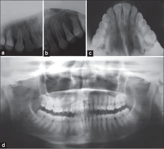
Figure 7: Posttreatment radiographs. (a) Intra oral periapical (IOPA) X-ray showed well aligned transposed maxillary right canine and lateral incisor. (b) IOPA X-ray showed well aligned impacted maxillary left canine. (c and d) Occlusal and orthopantomogram radiographs showed well-aligned transposed maxillary right canine and lateral incisor and impacted maxillary left canine