|


 |
| CASE REPORT |
|
| Year : 2012 | Volume
: 4
| Issue : 2 | Page : 110-111 |
|
|
A Rare Case of Spindle Cell Lipoma of Nose
Deepalakshmi Tanthry, PP Devan, Kavitha Ashok Kumar, Rukma Bhandary
Department of Otorhinolaryngology Head and Neck Surgery, AJ Institute of Medical Sciences, Kuntikana, Mangalore, Karnataka, India
| Date of Web Publication | 8-Apr-2013 |
Correspondence Address:
Deepalakshmi Tanthry
Department of Otorhinolaryngology Head and Neck Surgery, AJ Institute of Medical Sciences, Kuntikana, Mangalore, Karnataka
India
 Source of Support: None, Conflict of Interest: None
DOI: 10.4103/2006-8808.110250

 Abstract Abstract | | |
We present a case report of a 45-year-old lady with history of swelling on right side of the nose since two years. On clinical examination, there was a firm swelling, 3 × 2 cm in size, just above the right nasoalar crease, nontender and mobile. Computed tomography revealed fibrous tissue over anterior surface of the right maxilla and nasal bone with mild sclerosis of the right nasal bone. Excision was done through lateral rhinotomy incision. Histopathological examination of the excised specimen revealed spindle cell lipoma which is very rare. Very few cases have been reported in the literature so far. Keywords: Lateral rhinotomy, spindle cell lipoma, adipocyte tumor
How to cite this article:
Tanthry D, Devan P P, Kumar KA, Bhandary R. A Rare Case of Spindle Cell Lipoma of Nose. J Surg Tech Case Report 2012;4:110-1 |
How to cite this URL:
Tanthry D, Devan P P, Kumar KA, Bhandary R. A Rare Case of Spindle Cell Lipoma of Nose. J Surg Tech Case Report [serial online] 2012 [cited 2016 Jun 10];4:110-1. Available from: http://www.jstcr.org/text.asp?2012/4/2/110/110250 |
| Introduction | |  |
Spindle cell lipoma is a benign lipomatous tumor which constitutes about 1.5% of all adipocyte tumors. It was first described by Enzinger and Harvey in 1975. Similar to other kinds of lipomas, 75% of spindle cell lipomas are found in the subcutaneous tissue of back, shoulder, and neck. A spindle cell lipoma in face occurs infrequently. [1]
| Case Report | | |
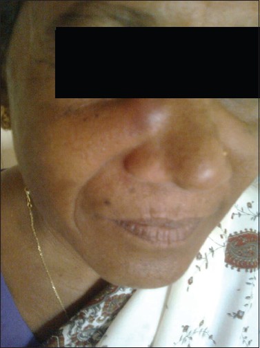
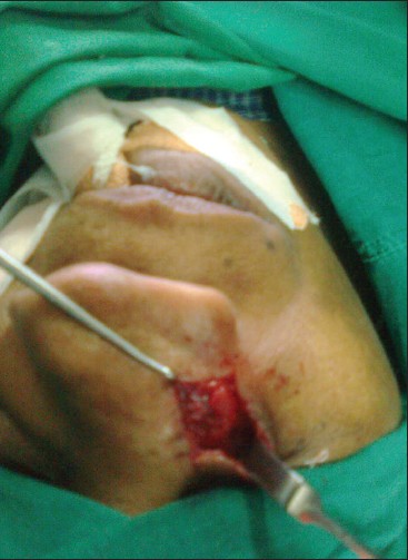
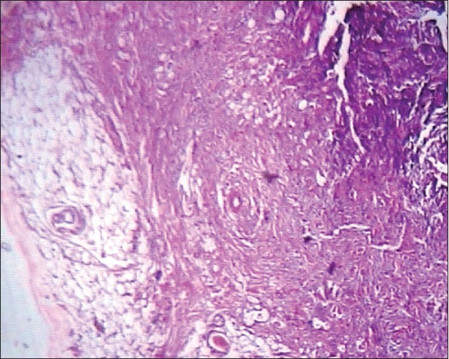
A 45-year-old lady presented to our outpatient department with a swelling in the right side of the nose since two years [Figure 1]. It was insidious in onset and gradually progressive. On clinical examination, there was a swelling 3×2 cm, 1 cm below medial canthus of the right eye extending to the right nasoalar crease. It was firm in consistency, nontender, and mobile. CT of the facial skeleton revealed a soft tissue mass over anterior surface of the right maxilla and right nasal bone with mild sclerosis of the right nasal bone. Excision was done under general anesthesia through lateral rhinotomy incision [Figure 2]. On gross examination, the mass was greyish red in color and firm in consistency. Incision was closed in layers and pressure dressing was applied. Specimen was sent for histopathological examination which revealed a mixture of mature adipocytes bland spindle cells in fibrous background with thick collagen bundles, consistent with spindle cell lipoma which is very rare in the region of head and face [Figure 3]. The patient was discharged after removal of the sutures. The patient was asymptomatic during the follow up.
| Discussion | | |
Spindle cell lipoma is a benign lipomatous tumor which usually arises on the back of the neck, shoulder, or upper back of males. [2] It constitutes about 1.5% of all adipose tissue neoplasms. Spindle cell lipomas are outnumbered by conventional lipomas by 60:1 in incidence. Angiolipoma, myelolipoma, spindle cell lipoma, chondrolipoma and myxolipoma are histological variants of lipomas arising from fat tissue. [3] Men are affected significantly more commonly than women (9:1) at a mean age in the sixth to seventh decade of life. [4] Our case is unusual because our patient was a female in fourth decade of her life. It is a subcutaneous tumor of back and shoulder usually solitary, subcutaneous, and well circumscribed. [5] It is relatively superficial with a mixture of mature adipocytes and bland spindle cells (pale eosinophilic cytoplasm with uniform wavy nuclei similar to neurofibroma) and multinucleated giant cells in mucinous or myxoid or fibrous background with thick collagen bundles. Spindle cells are arranged in short fascicles with occasional nuclear palisading. These tumors may have hemangiopericytic or angiomatous vascular pattern. They may have minimal or no fat. They may contain variable mast cell lymphocytes and characterized by abscence of storiform pattern, abscence of lipoblasts with no or rare mitotic activity. [6] These are associated with 13p and 16q abnormalities. The spindle cells stain positive for CD34 with androgen receptors in men and usually adipocytes stain positive for S-100. [7] The variable proportion of fibrous and myxoid elements among different example of these tumors confers to spindle cell lipoma a variable microscopic appearance that can make the diagnosis difficult. [8] These tumors do not have the tendency to recur. [5]
| Conclusion | | |
Spindle cell lipomas constitute only 7.5% of all adipocyte tumors, common in males in 6 th to 7 th decade of life in shoulder and upper back region. Our case report is mixture of mature adipocytes and bland spindle cells (pale eosinophilic cytoplasm with uniform wavy nuclei similar to neurofibroma) and multinucleated giant cells in mucinous or myxoid or fibrous background with thick collagen bundles. Spindle cells are arranged in short fascicles with occasional nuclear palisading unique in terms of rarity of its occurrence in a female of 45 years of age. This is one of a very few cases reported in literature so far.
| References | | |
| 1. | Monica M, Kibilda B, Gugala K. Spindle cell lipoma of the vestibule of nose. Otolaryngol Head Neck Surg 2008;139:25-6. 
|
| 2. | Mandal RV, Duncan LM, Austen WG, Nielsen GP. Infiltrating intramuscular spindle cell lipoma of face. J Cutan Pathol 2009;36:70-3.
|
| 3. | Fletcher CD, Martin BE. Spindle cell lipoma: A clinicopathological study with original observations. Histopathology 1987;11:803-17.
|
| 4. | Thompson LD. Spindle cell lipoma (Pathology). Ear Nose Throat J 2009;88:992-3.
|
| 5. | Piatlelli A, Rubini C, Fioroni M, Lezzi G. Spindle cell lipoma of cheek; A case report. Oral Oncol 2000;36:495-6.
|
| 6. | Patrick N, Lucas D. Soft tissue tumors pathology. Outlines.com.Inc; Available from: http://www.pathologyoutlines.com/topic/softtissueadiposepleomorphic.html [Last accessed on 2009].
|
| 7. | Wood L, Fountaine TJ, Rosamilia L, Helm KF, Clarke LE. Cutaneous CD34 cell lipomas: Histopathologic features distinguish spindle cell lipoma, solitary fibrous tumor, and dermatofibrosarcomaprotuberans. Am J Dermatopathol 2010;32:764-8.
|
| 8. | Diaz-Cascajo C, Borghi S, Weyers W. Fibrous spindle cell lipoma: Report of a new variant. Am J Dermatopathol 2001;23:112-5.
|
[Figure 1], [Figure 2], [Figure 3]
|