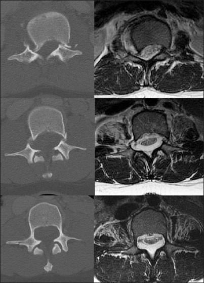
Figure 2: (Top) axial CT and T2 MRI at the level of L1 showing bilateral pedicle fractures and subluxation associated with canal compromise. (Middle) axial CT and T2 MRI at the level of L2 showing bilateral pedicle fractures and a dorsally located extradural meningeal cyst. (Bottom) axial CT and T2 MRI at the level of L3 showing aberrantly thin pedicles, anterior displacement of the neural elements, and dorsally located extradural meningeal cyst