
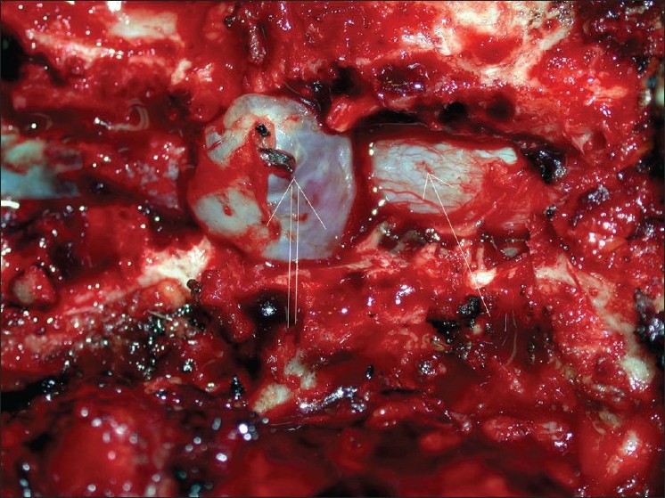
Figure 3: Intraoperative photo showing an extradural meningeal cyst (double arrow) dissected away from the dura of the thecal sac (single arrow)
| Close | |
|
|
|
|
Figure 3: Intraoperative photo showing an extradural meningeal cyst (double arrow) dissected away from the dura of the thecal sac (single arrow)
|
|