
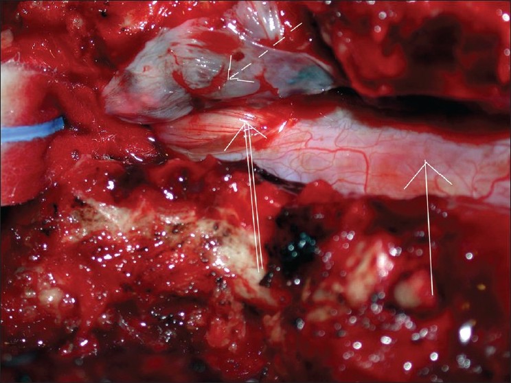
Figure 4: Intraoperative photo showing the emptied extradural meningeal cyst resected (broken arrow) and cyst ostium (double arrow) of the thecal sac (single arrow) laterally
| Close | |
|
|
|
|
Figure 4: Intraoperative photo showing the emptied extradural meningeal cyst resected (broken arrow) and cyst ostium (double arrow) of the thecal sac (single arrow) laterally
|
|