
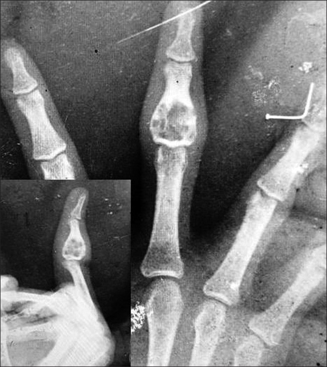
Figure 1: X‑ray showing a lytic, radiolucent, lobulated lesion with cortical expansion, a sclerotic rim, and septations at the base of the middle phalanx of the left middle finger. No calcification or periosteal reaction is noted
| Close | |
|
|
|
|
Figure 1: X‑ray showing a lytic, radiolucent, lobulated lesion with cortical expansion, a sclerotic rim, and septations at the base of the middle phalanx of the left middle finger. No calcification or periosteal reaction is noted
|
|