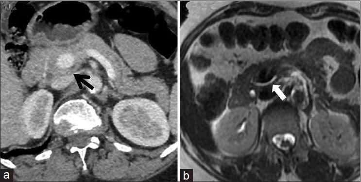
Figure 1: (a) Axial contrast-enhanced abdominal computed tomography scan displays a rind of pancreatic tissue encircling the portal vein consistent with portal annular pancreas. (b) Axial T2-weighted magnetic resonance imaging in addition displays the retroportal main pancreatic duct traversing posterior to the portal vein