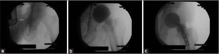
Figure 7: Intraoperative X‑rays demonstrating the adjustment of the femoral offset in a case of acetabulum osteomyelitis with subsequent septic hip joint arthritis. (a) The natural joint at the beginning of the surgery. (b) If a normal spacer is implanted, a decrease of the femoral offset is evident (notice the distance between the calcar and the os ischium). (c) By implanting a spacer according to the present technique, the femoral offset is increased