
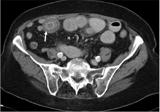
Figure 4: Axial CT image of the pelvis: There is a calculus (white arrow) identified within the small bowel lumen. Note the presence of both dilated and non‑dilated small bowel
| Close | |
|
|
|
|
Figure 4: Axial CT image of the pelvis: There is a calculus (white arrow) identified within the small bowel lumen. Note the presence of both dilated and non‑dilated small bowel
|
|