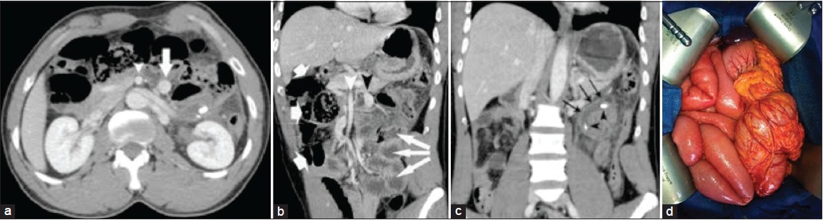
Figure 1: (a) Portal-venous phase computed tomography (CT) image. Superior mesenteric vein (arrow) is on the left side of the superior mesenteric artery (arrowhead); (b) Coronal multiplanar reformation (MPR) from portal-venous phase CT demonstrates a midgut malrotation. Superior mesenteric vein (black arrowhead) is on the left side of the superior mesenteric artery (white arrowhead). The entire small bowel (thin arrows) is in the left half of the abdomen and colon (thick arrows) is in the right half; (c) Coronal MPR from portal-venous phase CT demonstrates a acute upper left side appendicitis (arrows) with two stercoliths (arrowheads) and (d) Operative view with the entire small bowel in the right half of the abdomen and colon in the left half