
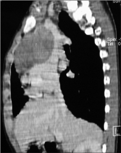
Figure 1: Computed tomography chest showing a large, lobulated, homogenous soft tissue anterior mediastinal mass (marked T). Fat planes with the superior vena cava, brachiocephalic vein and pericardium are maintained
| Close | |
|
|
|
|
Figure 1: Computed tomography chest showing a large, lobulated, homogenous soft tissue anterior mediastinal mass (marked T). Fat planes with the superior vena cava, brachiocephalic vein and pericardium are maintained
|
|