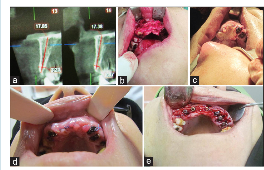
Figure 2: (a) Cone-beam computed tomography of anterior maxilla with thin width alveolar ridge. (b) Maxillary anterior buccal onlay bone grafting. (c) Tensionless coverage of bone graft with Anterior Palatal Island Advancement Flap. (d) Postoperative photograph 3 weeks after operation. (e) Implant insertion into the bone graft and adjacent fresh extraction socket