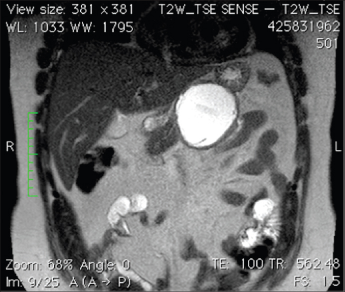
Figure 1: Coronal T2-weighted image of the pancreas showing a well-defined rounded cystic lesion abutting the ventral portion of the central pancreas. No daughter cysts could be seen indicating that the cyst is a Type CE 3 a (transitional hydatid cyst). The pancreatic duct is seen (as hyperintense line) leading to the lesion