|
 
 |
| CASE REPORT |
|
| Year : 2013 | Volume
: 15
| Issue : 1 | Page : 25-28 |
|
Herpes labialis after scaling and root planing: Related event or non-related event
CC Azodo1, P Erhabor2
1 Department of Periodontics, University of Benin, Benin-City, Nigeria
2 Department of Periodontics, University of Benin Teaching Hospital, Benin-City, Nigeria
| Date of Web Publication | 22-Sep-2014 |
Correspondence Address:
C C Azodo
Room 21, 2nd Floor, Department of Periodontics, Prof. Ejide Dental Complex, University of Benin Teaching Hospital, P.M.B. 1111 Ugbowo, Benin City, Edo State
Nigeria
 Source of Support: None, Conflict of Interest: None  | Check |
DOI: 10.4103/1595-1103.141390

Dental treatment may trigger the reactivation and multiplication of latent herpes virus in the trigeminal nerve ganglion, manifesting as herpes labialis. However, the reported dental treatment involved the use of local anesthetic agent either in form of infiltration or block. This article reported two cases of herpes labialis in otherwise healthy 63-year-old female and 40-year-old male after non-surgical periodontal treatment without local anesthesia using ultrasonic and manual scalers, respectively. They were not bothered by the condition and did not request for any specific care. However, warm saline mouthwash, analgesics, antibiotics, and lubricating cream for the angle of mouth without antiviral prescription were recommended. In conclusion, herpes labialis may be considered a potential post scaling and root planing complication of manual and ultrasonic methods after excluding other trigger factors. Authors hereby recommend the following: 1. Minimal chairside time for scaling and employment of adequate precaution geared toward minimizing trauma to the oral mucosa during scaling among younger practitioner. 2. Inclusion of herpes labialis as a complication of scaling and root planing to reduce chances of possible litigation. 3. Prescription of preventive medications 24 hours before dental treatment and continued for two days afterwards. Keywords: Herpes labialis, scaling and root planing, triggers
How to cite this article:
Azodo C C, Erhabor P. Herpes labialis after scaling and root planing: Related event or non-related event. Niger J Surg Res 2013;15:25-8 |
| Introduction | |  |
Herpes labialis , also known as fever blisters or cold sores, is a recurrent herpes simplex infection that usually affects the lips or the adjacent skin. It is one of the most prevalent and clinically obvious viral diseases presenting as bothersome, large, painful, and disfiguring lesions interfering with social activity and causing psychological problems. [1] It stands as one of most common infective vesiculo-ulcerative oral lesions with distressing and debilitating characteristics worldwide. Herpes labialis has been cited as a common cause of perioral discomfort that impairs appearance and quality of life. [2]
It is contagious for the previously uninfected individuals and those with compromised immune systems such as HIV-infected individuals and those undergoing chemotherapy. [3] Herpes labialis infection constitutes a serious risk to the dental team in the form of herpes whitlow and herpes keratitis during the treatment of patients with active lesions in the absence of proper infection control practices. [4]
Herpes labialis is a commonly occurring ailment with reported prevalence of 15-32.9%. [5],[6] It constitutes the third and fourth most prevalent oral mucosal lesion in children and youth in the USA [7] and in the adult population in Slovenia, respectively. [8] Herpes labialis has been reported to constitute 0.58% of oral mucosal lesions in patients visiting a dental school in Southern India. [9]
From initial manifestation to complete healing between 7-10 days, occasionally 14 days, it has five clinical stages: Prodromal, blister, weeping, scabbing, and healing. [10] The clinical diagnosis of herpes labialis is based on case-specific historical findings, characteristic clinical appearance, and the location of the lesions. [1] However, confirmatory laboratory diagnosis is necessary in form of viral culture, polymerase chain reaction, serology, direct fluorescent antibody testing, or Tzanck test. Prompt topical or oral antiviral therapy (acyclovir, valacyclovir, famciclovir, and penciclovir) is considerably effective in decreasing the severity and duration of herpetic episodes and contributes to the prevention of recurrence of herpes labialis. However, the value of antiviral therapy in the management of herpes labialis in immunocompetent patients remains a contentious issue. [11]
Literature review did not reveal any report of herpes labialis infection in patients after dental procedures without local anesthetic agents, such as scaling and root planing, which are non-surgical periodontal treatment procedures. We hereby report two cases of herpes labialis as possible complication or unrelated event after non-surgical periodontal treatment with ultrasonic and manual scalers.
| Case Report | | |
Case #1
A 63 year old female Edo State Ibo trader who is a known hypertensive and diabetic presented with a 3-day history of a sharp, continuous, and radiating pain in both upper and lower left quadrants. The pain was spontaneous at onset, associated with headache and sleep disturbance. There was history of gingival bleeding while brushing and self-perception of bad breath. She is a widow with seven children. She cleans her teeth once daily with a hard bristle brush and fluoride containing dentifrice.
On examination, plaque score was 2.33, calculus score 2.33, and oral hygiene was 4.66, which was poor oral hygiene status according to the Simplified Oral Hygiene Index. There was generalized chronic gingivitis, periodontal pocket depth of 5 mm on the distal aspect of 36, missing 46 as well as retained roots of 16. Periapical radiography revealed mild horizontal bone loss in 24, 25, 26, 27, 35, 36, and 37. Scaling and root planing of all quadrants of the mouth was done using ultrasonic scaler. This treatment was carried out by an intern under the supervision of a Consultant Periodontologist. Warm saline mouth wash was prescribed and oral hygiene instructions were given. Oral doses of amoxicillin [500 mg, 8 hourly for 5 days], metronidazole [400 mg, 8 hourly for 5 days], diclofenac sodium [50 mg, 12 hourly for 3 days], and vitamin C [100 mg, 8 hourly for 14 days] were also prescribed and given a 1-week appointment for review.
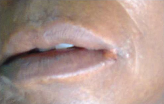
On recall visit, vesiculo-ulcerative lesion was seen on the left angle of the lips, which extended to the skin around the mouth and was consistent with the diagnosis of herpes labialis [Figure 1]. The patient was not bothered by the lesion but was informed of the self-resolving nature of the lesion. She was given another 1-week appointment for review. On second review, the herpes labialis had resolved and the patient was referred to Oral Surgery Clinic for extraction of the retained roots of the 1 st molar.
Case #2
A 40-year-old Urhobo man residing in Benin City, with previous history of uneventful tooth extraction, presented for scaling and polishing on advice of a relative. He is married with 3 children and works as a contract staff in a Federal Government establishment. He reported cleaning his teeth twice daily with a toothbrush and fluoride-containing toothpaste. He smokes and drinks alcohol occasionally. On examination plaque, score was 0.33, calculus score 2, and oral hygiene was 2.33, which is fair oral hygiene status according to the Simplified Oral Hygiene Index. The missing tooth was 47. Diagnosis of generalized chronic marginal gingivitis was made.
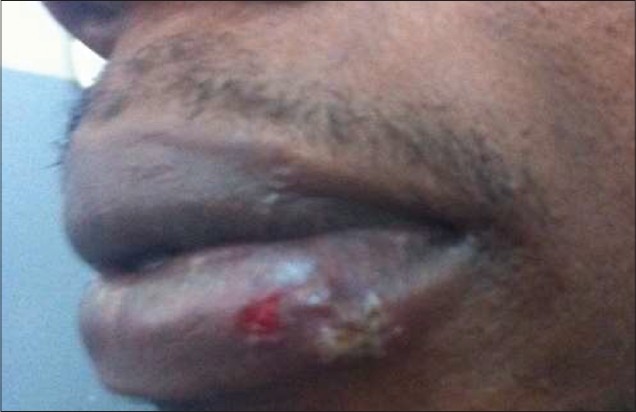
Scaling of whole mouth was done using manual scalers. This treatment was completed by a dental student under the supervision of a Senior Registrar and a Consultant Periodontologist. Three days after this scaling, patient noticed some rashes on his lips and associated generalized body pain [Figure 2]. The patient attributed it to malaria and started self-medication with artesunate. The patient presented to the clinic four days later with a vesiculo-ulcerative lesion on the lower lip, which was consistent with herpes labialis, and was advised on management of the lesion but was not bothered because his knowledge of self-resolving nature of the lesion.
| Discussion | | |
Herpes labialis occurs because herpes simplex virus has the unique property of becoming dormant after primary infection and subsequently reactivated following exposure to appropriate triggers. [12] Nikkels and Piθrard [13] reported herpes labialis following cosmetic procedures of the orofacial region, surgical and dental interventions, sun exposure, and burns. The scaling of calculus and removal of plaques using manual instruments puts pressure on the angle of the mouth while attending to the posterior teeth. This pressure can cause damage that may or may not be a visible trigger for herpes labialis. We speculate that during the procedure, the heat generated during ultrasonic scaling and pressure on angle of mouth during manual scaling may have caused injury unknown to the dentist and imperceptible to the patient, thus triggering the reactivation of the virus. Latrogenic trauma, which is more common in treatment rendered by young practitioners, has been implicated as a local trigger for herpes labialis. [13] In these two reported cases, the operators were a dental student and a house officer.
The poor and fair oral hygiene status in the two cases may have resulted in more chairside time in order to achieve thorough scaling. This may have been stressful and exhausting, thereby possibly serving as another form of trigger. Trauma, stress, fatigue, and iatrogenic injuries are among cited triggers for herpes labialis in the literature. [10],[11]
Although investigators have not fully elucidated the triggers that induce the virus to emerge from its dormancy to cause a recurrence, several factors have been identified: Ultraviolet light of both natural and artificial source (as in sunlight and tanning beds), lip chapping, lip trauma or abrasion, fever, menstruation, fatigue, overexposure to wind, extremes of temperature, immunosuppression, dental procedures, iatrogenic trauma, dietary factor, upper respiratory tract infection, digestive problems, traveler's diarrhea, decompression of the trigeminal nerve, pregnancy, and physical and emotional stress. Case #1 had diabetes mellitus, which will result in immunosuppression. Case #2 noted generalized body pain, which is commonly associated with malaria, in malaria endemic developing countries. Malaria causes fever and results in immunosuppression, both of which are established trigger factors for herpes labialis. [10],[11] When considering herpes labialis after dental procedure, it is therefore important to exclude malaria by self-reporting and laboratory investigation. The diagnosis in this report was based strictly on the history and clinical finding of oral lesions and no further investigation was done, which may be an obvious limitation.
It has been stated that herpes labialis is most prevalent in dental population of 21-40 years of age and a decreased prevalence with ageing was also noted. [13] In this case report, case #2 was 40 years, which was within the most prevalent age group while case #1 was 63 years.
It also appears that herpes labialis has higher tendencies to affect individuals of lower socioeconomic status and in this report, [12] both our patients were in the lower social class.
Studies have indicated that recurrent HSV lesions often develop 1-7 days after oral trauma, with the majority of lesions appearing 3-4 days after treatment. [14],[15] The observation in this report is therefore consistent with findings in documented literature. [14],[15]
| Conclusion | | |
Herpes labialis may be considered as a potentially post scaling and root planing complication of manual and ultrasonic methods after exclusion of other trigger factors. Authors recommend the following:
- Minimal chairside time for scaling and employment of adequate precaution geared toward minimizing trauma to the oral mucosa during scaling among younger practitioner
- Inclusion of herpes labialis as a complication of scaling and root planing to reduce chances of possible litigation
- The prescription of preventive medications 24 hours before dental treatment and continued for two days afterwards.
| References | | |
| 1. | Siegel MA. Diagnosis and management of recurrent herpes simplex infections. J Am Dent Assoc 2002;133:1245-9. 
[PUBMED] |
| 2. | Arduino PG, Porter SR. Oral and perioral herpes simplex virus type 1 (HSV-1) infection: Review of its management. Oral Dis 2006;12:254-70.
|
| 3. | Sparano JA, Sarta C. Infection prophylaxis and antiretroviral therapy in patients with HIV infection and malignancy. Curr Opin Oncol 1996;8:392-9.
|
| 4. | Browning WD, McCarthy JP. A case series: Herpes simplex virus as an occupational hazard. J Esthet Restor Dent 2012;24:61-6.
|
| 5. | Embil JA, Stephens RG, Manuel FR. Prevalence of recurrent herpes labialis and aphthous ulcers among young adults on six continents. Can Med Assoc J 1975;113:627-30.
[PUBMED] |
| 6. | Young TB, Rimm EB, D'Alessio DJ. Cross-sectional study of recurrent herpes labialis. Prevalence and risk factors. Am J Epidemiol 1988;127:612-25.
|
| 7. | Shulman JD. Prevalence of oral mucosal lesions in children and youths in the USA. Int J Paediatr Dent 2005;15:89-97.
[PUBMED] |
| 8. | Kovac-Kovacic M, Skaleric U. The prevalence of oral mucosal lesions in a population in Ljubljana, Slovenia. J Oral Pathol Med 2000;29:331-5.
|
| 9. | Mathew AL, Pai KM, Sholapurkar AA, Vengal M. The prevalence of oral mucosal lesions in patients visiting a dental school in Southern India. Indian J Dent Res 2008;19:99-103.
[PUBMED]  |
| 10. | Sciubba JJ. Herpes simplex and aphthous ulcerations: Presentation, diagnosis and management--an update. Gen Dent 2003;51:510-6.
[PUBMED] |
| 11. | Straus SE, Rooney JF, Sever JL, Seidlin M, Nusinoff-Lehrman S, Cremer K. NIH Conference. Herpes simplex virus infection: Biology, treatment, and prevention. Ann Intern Med 1985;103:404-19.
[PUBMED] |
| 12. | Crivelli MR, Aguas S, Adler I, Quarracino C, Bazerque P. Influence of socioeconomic status on oral mucosa lesion prevalence in schoolchildren. Community Dent Oral Epidemiol 1988;16:58-60.
|
| 13. | Nikkels AF, Pièrard GE. Treatment of mucocutaneous presentations of herpes simplex virus infections. Am J Clin Dermatol 2002;3:475-87.
|
| 14. | Kriesel JD, Pisani PL, McKeough MB, Baringer JR, Spruance SL. Correlation between detection of herpes simplex virus in oral secretions by PCR and susceptibility to experimental UV radiation-induced herpes labialis. J Clin Microbiol 1994;32:3088-90.
|
| 15. | Spruance SL, Kriesel JD, Evans TG, McKeough MB. Susceptibility to herpes labialis following multiple experimental exposures to ultraviolet radiation. Antiviral Res 1995;28:57-67.
|
[Figure 1], [Figure 2]
|


 Search Pubmed for