|
 
 |
| CASE REPORT |
|
| Year : 2016 | Volume
: 11
| Issue : 1 | Page : 23-25 |
|
Hydatid cyst of the pancreas: A case report in the West of Iran
Mazaher Ramezani1, Hadi Yarahmadi1, Masoud Sadeghi2
1 Molecular Pathology Research Center, Emam Reza University Hospital, Kermanshah University of Medical Sciences, Kermanshah, Iran
2 Cancer Research Center, Kermanshah University of Medical Sciences, Kermanshah, Iran
| Date of Web Publication | 10-Mar-2016 |
Correspondence Address:
Masoud Sadeghi
Cancer Research Center, Kermanshah University of Medical Sciences, Kermanshah
Iran
  | Check |
DOI: 10.4103/1858-5000.178509 
Echinococcus granulosus is caused hydatid cyst disease that is a common parasitic disease in endemic region. In this study, we report our experience with a case of hydatid cyst involving the pancreas in the West of Iran. A 65-year-old woman admitted to our hospital with abdominal discomfort presenting for 4 months. Computed tomography scan revealed mass measuring 5 cm × 4 cm with increased wall thickness in the head of the pancreas. A cystic adenocarcinoma was included in the differential diagnosis. Tumor markers (carbohydrate antigen 19-9, carcinoembryonic antigen, and alpha-fetoprotein) had normal range. On abdominal exploration, whipple surgery was done. Histopathologic findings revealed a well-defined creamy-whitish mass measuring 6 cm × 4 cm × 3 cm attached to small intestine. The microscopic examination revealed a typical hydatid cyst which contained some scolices. After this case, we believe hydatid disease must be considered in the differential diagnosis of pancreatic cysts, especially in countries where echinococcosis is endemic. In this condition, use of specific antigenic tests can be helpful for preoperative diagnosis and appropriate management of the disease. Keywords: Case report, hydatid cyst, pancreas, Western Iran
How to cite this article:
Ramezani M, Yarahmadi H, Sadeghi M. Hydatid cyst of the pancreas: A case report in the West of Iran. Sudan Med Monit 2016;11:23-5 |
| Introduction | |  |
Echinococcus granulosus, also called the hydatid worm, is caused hydatid cyst disease that is a common parasitic disease in the endemic region. The eggs of the worm being excreted in the feces of infected dogs. [1],[2] Intermediate hosts are usually cows, sheeps, and pigs, whereas human beings are accidental intermediate hosts. After ingestion, eggs hatch in the jejunum. Larvae enter the portal system through intestinal mucosa. [3] It may found in any organ of the body, but liver is the more common location. Hydatidosis of other organ such as the brain, kidney, bone, heart, and pancreas is rare. [4] Even in the endemic countries, involvement of pancreas it has been reported in only 0.1-2% of patient with hydatid disease. [1],[3],[5] The diagnosis before surgery is difficult because the presenting symptoms and radiological finding may be similar to other common cystic lesions of the pancreas and thus must be confirmed by histopathology. In this study, we report our experience with a case of hydatid cyst involving the pancreas in the West of Iran.
| Case Report | | |
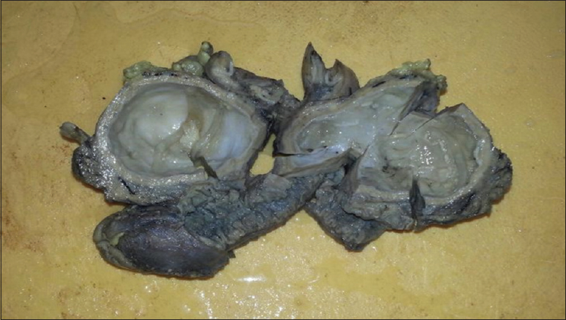
A 65-year-old woman admitted to our hospital with abdominal discomfort presenting for 4 months. Abdominal sonography suggested a septated cyst measuring (52 mm) adjacent to the right lobe of liver and posteromedial location of gallbladder. Computed tomography (CT) scan revealed mass measuring 5 cm × 4 cm with an increased wall thickness in the head of the pancreas. A cystic adenocarcinoma was included in the differential diagnosis. On laboratory investigation, level of amylase and lipase increased and slowly decreased after operation. Tumor markers (carbohydrate antigen 19-9, carcinoembryonic antigen, and alpha-fetoprotein) had normal range. On abdominal exploration, whipple surgery was done. Histopathologic findings revealed a well-defined creamy-whitish mass measuring 6 cm × 4 cm × 3 cm attached to small intestine. On opening the sample, a multiloculated cyst was seen which contained clear fluid and wall thickness was 5 mm [Figure 1]. The microscopic examination revealed a typical hydatid cyst which contained some scolices [Figure 2]a and b. | Figure 2: Scolices of echinococcus, Hematoxylin and Eosin staining (a) ×10 magnification (b) ×40 magnification
Click here to view |
| Discussion | | |
Liver and lung are involved by human hydatidosis in 85-95% of cases and other organs involvement is about 5-15% of the cases. [6] Primary hydatid cysts of the pancreas are rare, it must be considered in the differential diagnosis of pancreatic tumor with cystic components particularly in endemic regions for disease. Clinical presentation of hydatid disease of the pancreas is the result of pressure by the cyst on adjacent structures. Signs and symptoms depend on the size and anatomical location of the cyst. The location of the hydatid cyst in the pancreas is variable. [5],[7]
Hydatid cyst of head, it is usually discovered during a complication, it may cause obstructive Jaundice, acute pancreatitis, and recurrent acute or chronic pancreatitis. Cyst located in body and tail can be asymptomatic or can present as only an abdominal mass. Portal hypertension is also a manifestation of pancreatic hydatid cyst. [6] The head of the pancreas is the most frequent location involved by hydatid disease. [8]
Primary pancreatic hydatid cysts are difficult to diagnose preoperatively. The presence of cystic lesions of pancreas is easily identified by ultrasound, CT scan, and magnetic resonance imaging, but not specific. Serological tests are positive. Tests are positive in up to 80% of abdominal hydatid cysts. [6],[8] The enzyme-linked immunosorbent assay test for echinococcal antigens is positive in over 85% of infected patients. [3] As the results of preoperative imaging studies did not bring hydatid disease into consideration that we did not perform these tests preoperatively. [9] It has been recommended to obtain a fine needle aspiration biopsy for definite diagnosis and for appropriate treatment planning. [10]
| Conclusions | | |
After this case, we believe hydatid disease must be considered in the differential diagnosis of pancreatic cysts, especially in countries where echinococcosis is endemic. In this condition, use of specific antigenic tests can be helpful for preoperative diagnosis and appropriate management of the disease.
Financial support and sponsorship
Nil.
Conflicts of interest
There are no conflicts of interest.
| References | | |
| 1. | Unal B, Elpek GO, Yildirim S, Gelen T, Erdogan O, Ozkaynak C. Echinococcus multilocularis infestation in the head of the pancreas. J Clin Exp Pathol 2014;4:167.  |
| 2. | Bansal VK, Misra MC, Krishna A, Kumar S, Garg P, Khan RN, et al. Pancreatic hydatid cyst masquerading as cystic neoplasm of pancreas. Trop Gastroenterol 2010;31:335-7. |
| 3. | Khoshmohabat H, Moqadam JA, Sohrabi SZ, Kalantar Motamedi MH, Azizi T. Hydatid cyst of the pancreas mimicking neoplasm. J Coll Physicians Surg Pak 2014;24 Suppl 2:S84-5. |
| 4. | Baghbanian M, Salmanroghani H, Karegar S, Binesh F, Baghbanian A. Pancreatic tail hydatid cyst as a rare cause for severe acute pancreatitis: A case report. Govaresh 2013;18:57-61. |
| 5. | Moosavi SR, Kermany HK. Epigastric mass due to a hydatid cyst of the pancreas. A case report and review of the literature. JOP 2007;8:232-4. |
| 6. | Trigui A, Rejab H, Guirat A, Mizouni A, Ben Amar M, Mzali R, et al. Hydatid cyst of the pancreas. About 12 cases. Ann Ital Chir 2013;84:165-70. [ PUBMED] |
| 7. | Masoodi MI, Nabi G, Kumar R, Lone MA, Khan BA, Naseer Al Sayari K. Hydatid cyst of the pancreas: A case report and brief review. Turk J Gastroenterol 2011;22:430-2. |
| 8. | Ousadden A, Elbouhaddouti H, Ibnmajdoub KH, Mazaz K, Aittaleb K. Primary hydatid cyst of the pancreas with a hepatic pedicule compression. Cases J 2009;2:201. |
| 9. | Ozmen MM, Moran M, Karakahya M, Coskun F. Recurrent acute pancreatitis due to a hydatid cyst of the pancreatic head: A case report and review of the literature. JOP 2005;6:354-8. |
| 10. | Ugras S, Sakarya ME, Arslan H, Bozkurt M, Akdeniz H. The diagnosis by fine needle aspiration biopsy of hydatid cyst of the pancreas. Acta Chir Belg 1997;97:244-6. |
[Figure 1], [Figure 2]
|


 Search Pubmed for