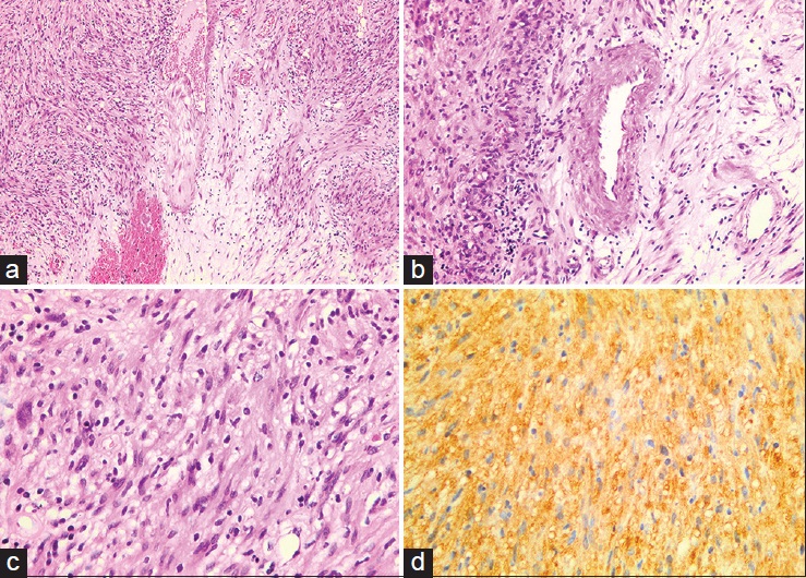
Figure 2: (a) Spindle cell proliferation in hypocellular and hypercellular zonation (×10 objective) (b) Thick walled vascular spaces and vague palisading of tumour cells (×20 objective) (c) The cells show random mild cytological atypia. No increased mitotic activity or necrosis (×40 objective) (d) S100 shows diffuse positivity (×40 objective)