- Institution: Stanford Univ Med Ctr Lane Med Lib/Periodical Dept/Rm L109
- Sign In as Member / Individual
BLOOD-BRAIN BARRIER DRUG TARGETING: THE FUTURE OF BRAIN DRUG DEVELOPMENT
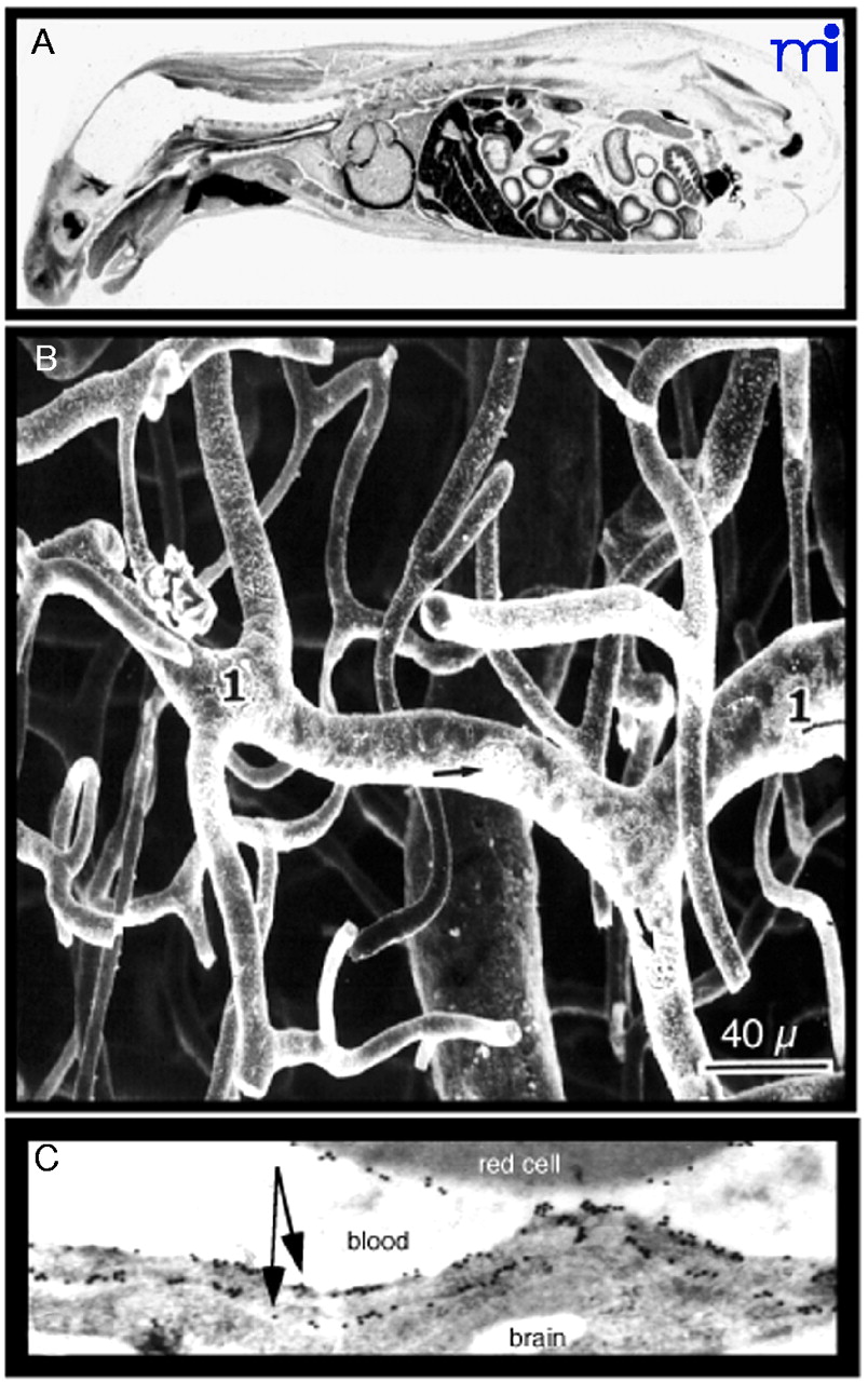
Structure of the Blood–Brain Barrier (BBB). A.
Autoradiograph of a mouse taken 30 minutes after intravenous injection of radiolabeled histamine, a small molecule that does not cross the blood–brain barrier. The drug is taken up by all organs of the body except the brain and spinal cord. Reprinted with permission (6). B. Scanning electron micrograph of the vascular cast of the human cortical microvasculature. The capillaries are separated by a distance of approximately 40 μm. Thus, each neuron is virtually perfused by its own blood vessel. Reprinted with permission (10). C. Immunogold electron micrograph of the capillary endothelium of the human brain stained with an antibody specific for the GLUT1 glucose transporter. The transporter is found on the erythrocyte plasma membrane, on the luminal membrane of the capillary endothelium, which is the blood side of the BBB, and on the abluminal membrane of the capillary endothelium, which is the brain side of the BBB. A distance of approximately 300 nm separates the luminal and abluminal membranes of the capillary endothelium (arrows). Reprinted with permission (59).

