- Institution: Stanford Univ Med Ctr Lane Med Lib/Periodical Dept/Rm L109
- Sign In as Member / Individual
BLOOD-BRAIN BARRIER DRUG TARGETING: THE FUTURE OF BRAIN DRUG DEVELOPMENT
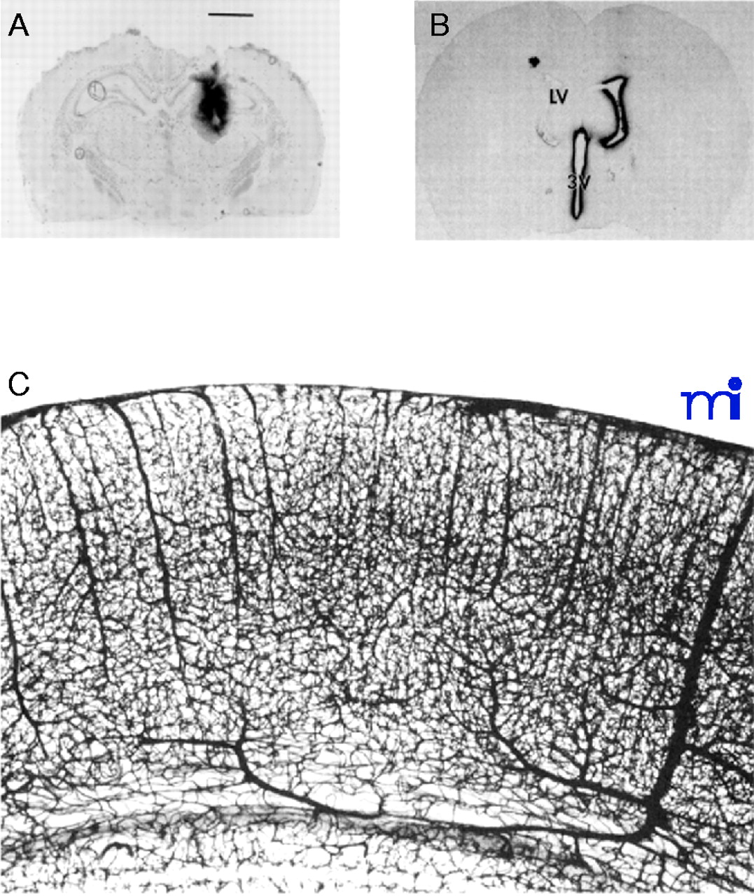
Transcranial and transvascular drug delivery to the the brain. A. Film autoradiogram of rat brain 48 hours after an intracerebral implantation of a polymeric disc containing radiolabeled nerve growth factor (NGF). The magnification bar is 2.5 mm, and the diameter of the polymeric implant is 2.0 mm. Therefore, there has been little distribution of the drug away from the polymeric implant during the 48-hour period. Reprinted with permission (7). B. Autoradiograph of rat brain 20 hours after a single intracerebroventricular injection of radiolabeled brain derived neurotrophic factor (BDNF) into the lateral ventricle (LV). The drug distributes only to the ependymal surface of the ipsilateral lateral ventricle and to the third ventricle (3V) prior to exodus from the spinal fluid compartment back to the peripheral bloodstream. There is no significant distribution of the drug to the contralateral side, and there is no significant penetration of the drug into brain parenchyma from the ependymal surface. Reprinted with permission (8). C. India ink injection study of rat brain showing the density of the cortical microvasculature. Because brain capillaries are separated by a distance of only about 40 microns, any drug that crosses the vascular barrier via the transvascular route to brain will immediately distribute to the extracellular space of the entire brain. Reprinted with permission (9).

