- Institution: Stanford Univ Med Ctr Lane Med Lib/Periodical Dept/Rm L109
- Sign In as Member / Individual
Reciprocal Interaction of Sleep and Synaptic Plasticity
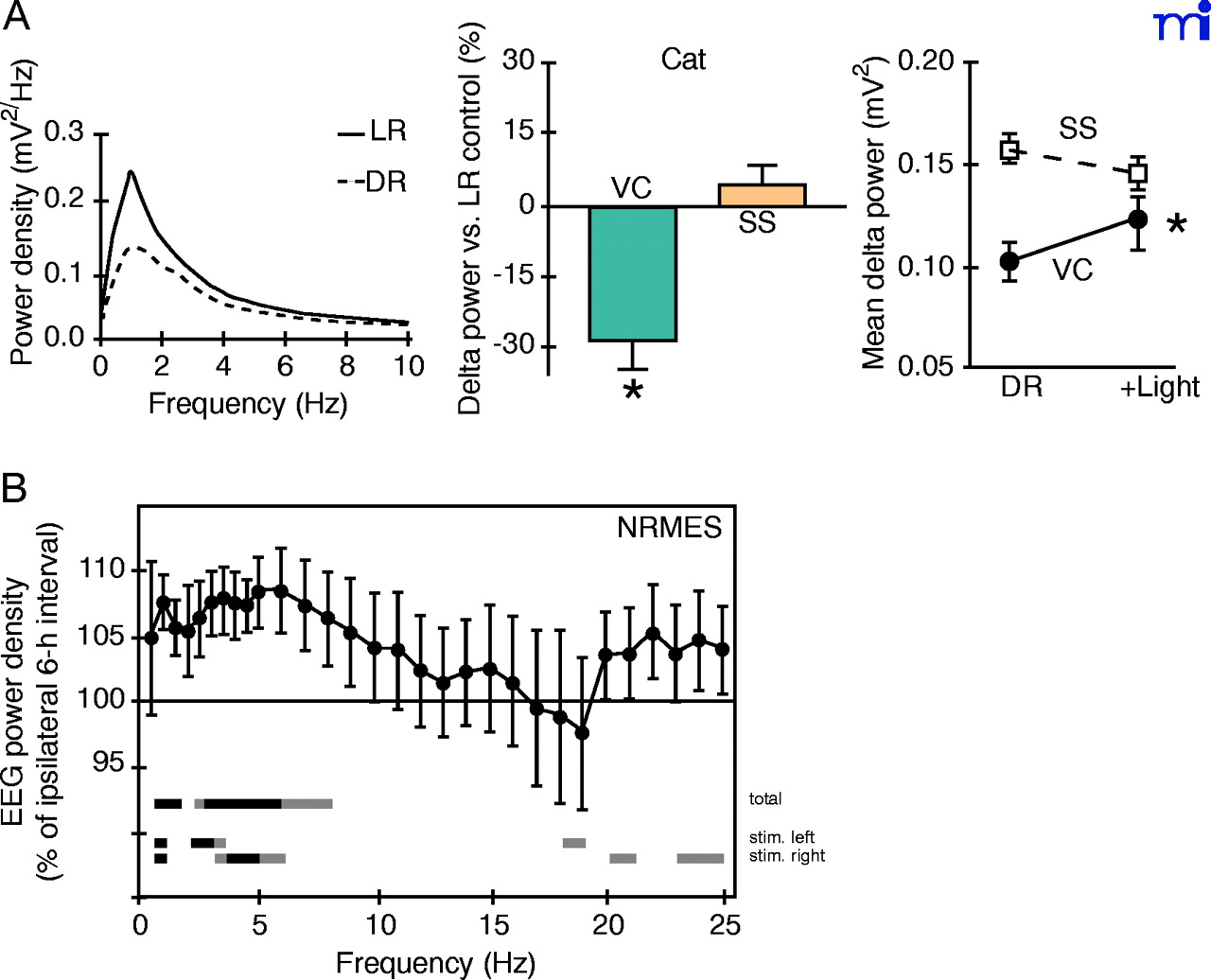
Sensory experience affects quality of subsequent sleep. A. Early sensory deprivation reduces slow-wave activity. (Left) Representative SWS power spectra of light-reared (LR) and dark-reared (DR) cats. DR kittens were raised in complete darkness from birth for 4 months. (Center) Group data. Reduction of delta power after DR is restricted to visual cortex (VC) and not seen in somatosensory area (SS). (Right) Slow-wave plasticity is reversible. Recovery of delta activity after light re-exposure for one to two months. Adapted from Miyamoto et al. (53). B. Sensory stimulation during waking enhances interhemispheric asymmetry of EEG power density. Ordinate indicates EEG power density of hemisphere contralateral to intact whisker side as a percentage of ipsilateral hemisphere (whisker-cut side) during SWS. Lines above the abscissa indicate significant difference between hemispheres. Top line (total) represents statistics for all data, middle and bottom lines for stimulation of intact left or right side whiskers. Adapted from Vyazovski et al. (54).

