- Institution: Stanford Univ Med Ctr Lane Med Lib/Periodical Dept/Rm L109
- Sign In as Member / Individual
LIPID Arrays: New Tools in the Understanding of Membrane Dynamics and Lipid Signaling
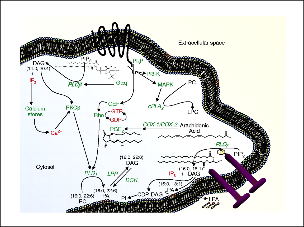
Lipid signaling pathways in mammalian cells. Some of the richness and complexity of lipid signaling is conveyed by the conventional designations shown above: DAG, for instance, is not a single species, but rather a variety of diglycerides with varying acyl chains. Distinct cell signaling pathways may mediate the generation of specific lipid products through the use of differential isoenzymes (e.g., PLCβvs PLCγ). Similarly, DGKεhas a preference for 20:4 DAG as a substrate. Lipidomic approaches identify distinct lipid species, such as specific acyl species of DAG, according to their individual roles in signaling. In other words, not all DAGs may be created equally nor have the same effects on the cell. Further elucidation of the signaling pathways shown above into discrete reaction mechanisms will facilitate understanding the roles of lipids in cellular processes. Proteins are indicated in green text, lipid species in black text, and other small molecules in red text; a GPCR is shown as a black band with seven transmembrane domains; a pair of nail-like geometries represents an active receptor tyrosine kinase (lower right). (PC, phosphatidylcholine; PI, phosphatidylinositol; PA, phosphatidic acid; DAG, diacylglycerol; LPA, lysophosphatidic acid; LPC, lysophosphatidylcholine; IP3, inositol 1,4,5-trisphosphate; PIP2, phosphatidylinositol bisphosphate; PIP3, phosphatidylinositol trisphosphate; PLC, phospholipase C; PLD, phospholipase D; PLA, phospholipase A; LPP, lipid phosphate phosphatase; DGK, diacylglycerol kinase; GEF, guanine nucleotide exchange factor; PKC, protein kinase C; PI3-K, phosphatidylinositol 3-kinase; MAPK, mitogen activated protein kinase; COX, cyclooxygenase; PGE2, prostaglandin E2; CDP-DAG, cytidine diphosphate-diacylglycerol.)

