- Institution: Stanford Univ Med Ctr Lane Med Lib/Periodical Dept/Rm L109
- Sign In as Member / Individual
Fibrin Mechanisms and Functions in Nervous System Pathology
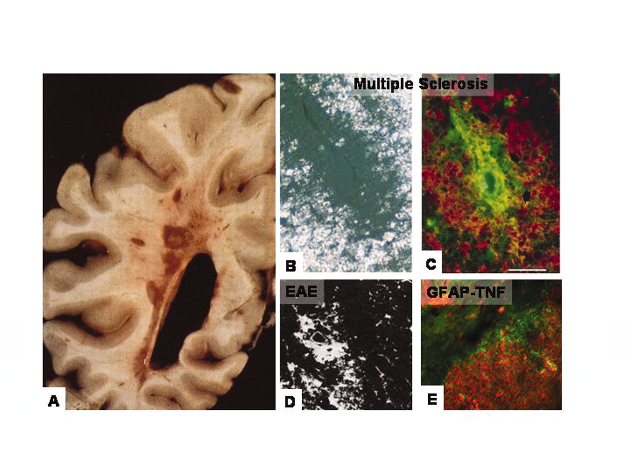
Fibrin deposition in inflammatory demyelinating plaques of MS and respective animal models. A. Unstained gross coronal section of brain shows typical, slightly hemorrhagic, periventricular, and parenchymatous lesions. MS plaque viewed under polarized light (B) and double labeled for fibrinogen (green) and macrophages (red) (C). High macrophage density is associated with fibrinogen leakage. Two rodent models for MS also reveal fibrin depositions in inflammatory demyelination. D. In EAE, foci of fibrin deposits are observed perivascularly. E. In a TNF-transgenic mouse model for MS, fibrin deposition (red) is present in regions of macrophage infiltration (green). From (80); Brain 120, Gay et al., “The application of multifactorial cluster analysis in the staging of plaques in early multiple sclerosis. Identification and characterization of the primary demyelinating lesion,” 1461–1483 (1997), by permission of Oxford University Press. From (98); Fed. Proc. 35, Paterson, “Experimental allergic encephalomyelitis: Role of fibrin deposition in immunopathogenesis of inflammation in rats,” 2428–2434 (1976), by permission from FASEB. From (146); Poser, An Atlas of Multiple Sclerosis. The Parthenon Publishing Group Inc, New York, NY, p. 60 (1998), by permission of CRC Press.

