- Institution: Stanford Univ Med Ctr Lane Med Lib/Periodical Dept/Rm L109
- Sign In as Member / Individual
RHO SIGNALING in Vascular Diseases
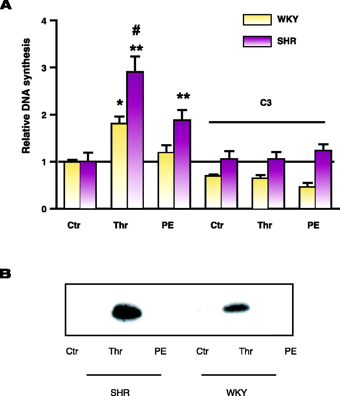
Elevated DNA synthesis and RhoA activation in spontaneously hypertensive rats. A. DNA synthesis in aortic smooth muscle cells from spontaneously hypertensive rats (SHR) and normotensive Wistar-Kyoto (WKY) rats. Cells were grown to confluence and serum-starved for 24 hours and then exposed to either vehicle, thrombin (Thr,12nM) or phenylephrine (PE, 10 μM) for 48 hours and labeled with [3H]thymidine for the last five hours prior to harvest for measurement of [3 H]thymidine incorporation into DNA. [See (45).] B. RhoA activation in aortic smooth muscle cells from SHR and WKY rats is measured as outlined in the text. Cells were stimulated with vehicle, thrombin (Thr,12nM) or phenylephrine (PE, 10μM) for 3 minutes. Cells were lysed and the lysates centrifuged at 14,000 x g for two minutes. The remaining supernatant was either used for Western blot analysis for total RhoA or added to a GST-fusion protein of the Rho binding domain of the Rho effector rhotekin precoupled to glutathione beads, followed by Western blot analysis for active RhoA.

