- Institution: Stanford Univ Med Ctr Lane Med Lib/Periodical Dept/Rm L109
- Sign In as Member / Individual
Twenty Years of Calcium Imaging: Cell Physiology to Dye For
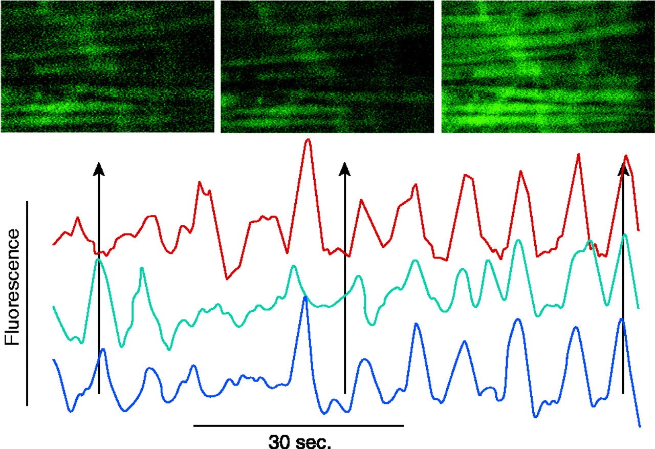
Figure 2.
Confocal image of a rat mesenteric small artery loaded with a Ca2+-sensitive dye. The horizontal streaks are smooth muscle cells with an overlaying network of sympathetic nerve fibers. The graph shows a time series of three smooth muscle cells. The Ca2+ oscillations are first unsynchronized and later synchronized. The scale bar is 4 μm. (Image kindly provided by Christian Aalkjaer, University of Aarhus, Denmark.)

