- Institution: Stanford Univ Med Ctr Lane Med Lib/Periodical Dept/Rm L109
- Sign In as Member / Individual
Caenorhabditis elegans in Parkinson’s Disease Drug Discovery: Addressing an Unmet Medical Need
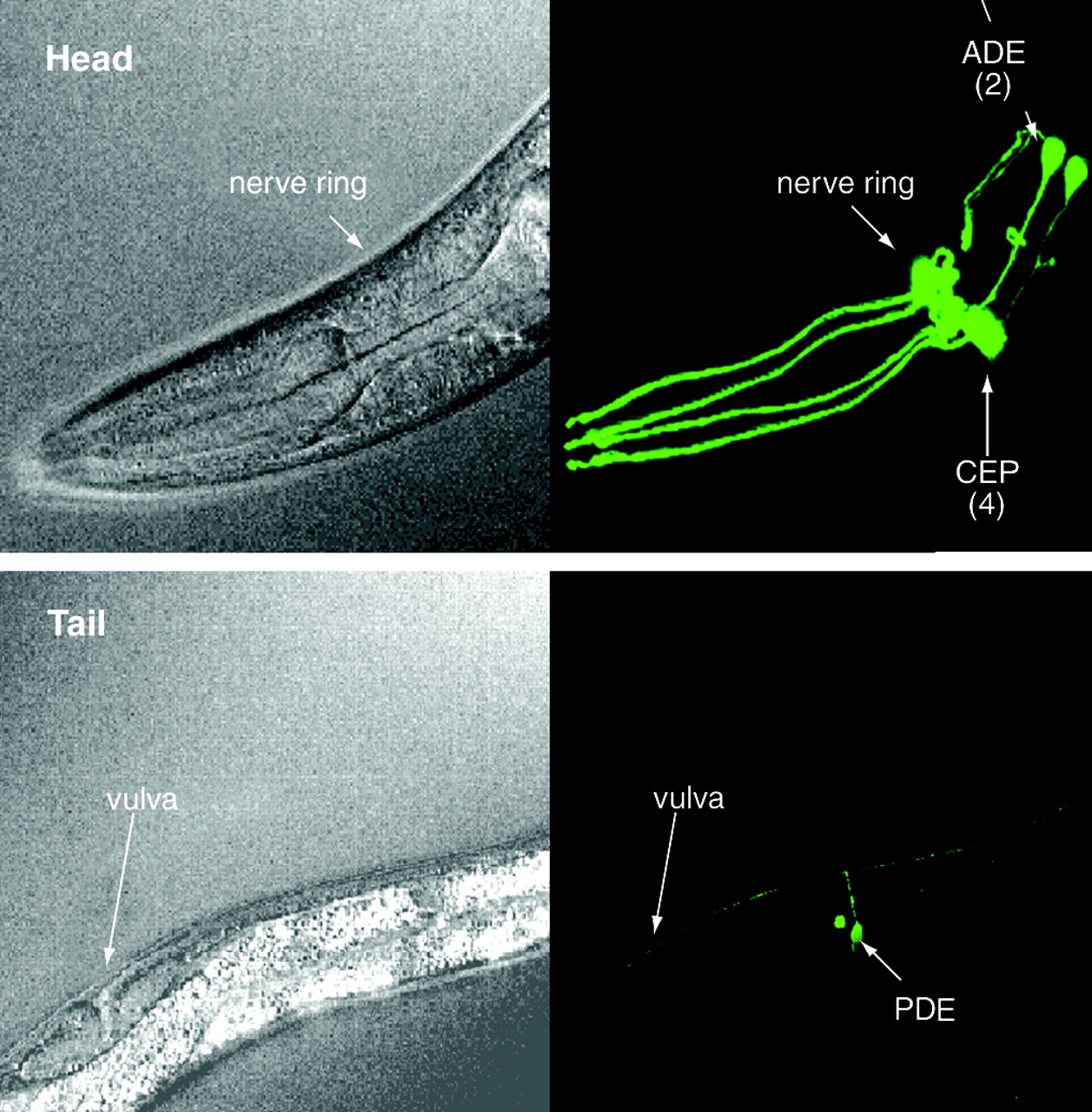
Dopaminergic neuroanatomy in theC. elegans hermaphrodite. Adult wild-type worms contain eight dopamine neurons, which can be visualized in transgenic animals that express the green fluorescent protein (see text for details of dopaminergic neuron–specific expression of GFP). The head of the animal (upper panel; the pharynx is labeled for visual orientation) includes two anterior deirid (ADE) neurons and four cephalic sensilla (CEP) neurons; arrows point to cell bodies. The tail of the animal (lower panel; the vulva is labeled) includes two posterior deirid (PDE) neurons; arrows point to cell bodies. Live animals were imaged through differential interference contrast microscopy (left images) and confocal epifluorescence microscopy (right images). Reproduced with permission from (26).

