- Institution: Stanford Univ Med Ctr Lane Med Lib/Periodical Dept/Rm L109
- Sign In as Member / Individual
Antifibrillatory Agents and Potassium Channels in the Atria: Pore Block versus Channel Trafficking
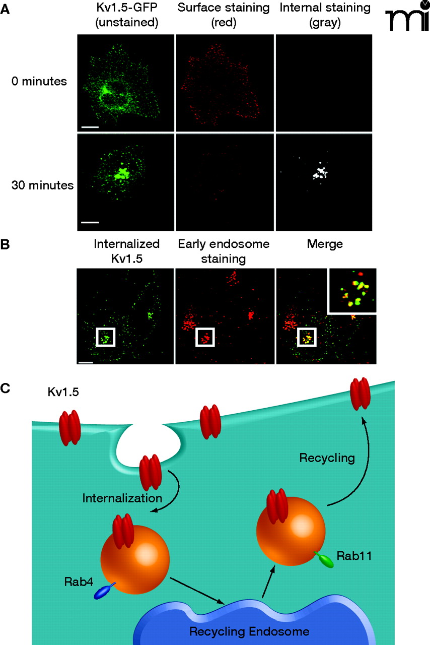
Trafficking of the Kv1.5 channel in HL-1 atrial myocytes. The Kv1.5 channel was constructed as a green fluorescence protein–fusion product in which a single GFP is inserted into an extracellular domain of Kv1.5; the GFP sequence functions as a single epitope in the immunostaining of expressed Kv1.5–GFP described here. (All scale bars are 5 μm.) A. Internalization of Kv1.5–GFP can be monitored as redistribution of green fluorescence over time [left panel; cells following 0 (top) and 30 minutes (bottom) incubation at 37oC are respectively shown]. Because the cells shown were incubated in the presence of a non-fluorescing primary anti-GFP antibody that does not affect trafficking, co-incubation of the cells at 0 or 30 minutes with a saturating red-fluorescing secondary antibody (middle panel) indicates the population of Kv1.5–GFP that remains accessible at the cell surface at the given time; subsequent fixation, permeabilization, and incubation of the cells with a gray-appearing antibody (i.e., non-red; right panel) identifies Kv1.5–GFP that has been internalized from the cell surface. B. Confocal imagery establishes that internalized Kv1.5 can be trafficked to early endosomes. The internalization assay was performed for 30 minutes as described above. Cells were fixed, permeabilized, and stained for internalized Kv1.5 as well as for the early endosomal marker EEA1. The merged image shows co-localized of channel with EEA1 as indicated by yellow punctae. C. The schematic represents the Kv1.5 internalization and recycling pathway. As verified by the confocal imagery with internalized Kv1.5 and shown in the schematic, trafficking of Kv1.5 is both Rab4- and Rab11-dependent. (See text for details.)

