- Institution: Stanford Univ Med Ctr Lane Med Lib/Periodical Dept/Rm L109
- Sign In as Member / Individual
The Biology of Caveolae: Lessons from Caveolin Knockout Mice and Implications for Human Disease
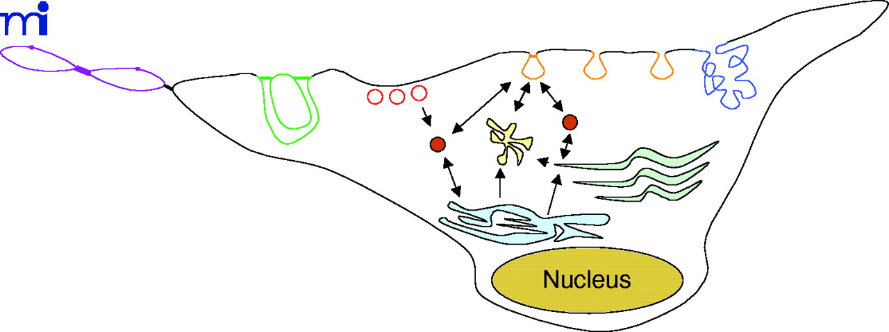
Figure 3.
Intracellular trafficking of caveolae-related compartments. The following subcellular membrane compartments are shown: fenestrae (violet), channels (green), plasmalemma vesicles (red), caveolae (orange), vesiculo-vacuolar organelles (blue), caveosomes (yellow), and cavicles (solid rust circles). Arrows depict the movement of caveolae-derived vesicles (orange) from the cell surface to caveosomes (yellow), the Golgi apparatus (light green), and the ER (light blue).

