- Institution: Stanford Univ Med Ctr Lane Med Lib/Periodical Dept/Rm L109
- Sign In as Member / Individual
Ceramide-1-phosphate: The “missing” link in eicosanoid biosynthesis and inflammation

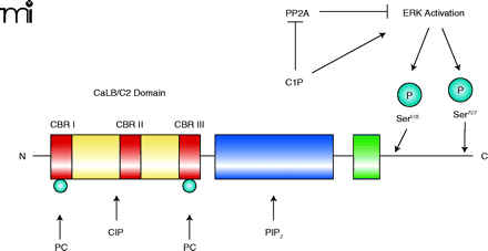
A hypothetical mechanism of cPLA2activation in response to the generation of C1P. The structure of cPLA2α includes a CaLB/C2 domain (red and yellow), containing calcium binding regions (CBRs I, II and III; red), the PIP2 domain (i.e., the catalytic domain; blue), and the hinge region (green). C1P has multiple roles in activating cPLA2α. Within the structure of cPLA2α, Ser505 and Ser727 can be phosphorylated (blue spheres), by ERK, to activate cPLA2α; C1P is shown as a direct activator of ERK as well as an indirect activator of ERK by inhibiting protein phosphatase 2A (PP2A). C1P also interacts directly with cPLA2 near CBRII. Additionally, C1P increases calcium (Ca2+) concentrations, in a compartmentalized manner, in order to induce the association of cPLA2α with perinuclear membranes. Sites of interaction with C1P and phosphatidylcholine (PC) are also indicated. See text for details.


