- Institution: Stanford Univ Med Ctr Lane Med Lib/Periodical Dept/Rm L109
- Sign In as Member / Individual
CALCIUM: A Role for Neuroprotection and Sustained Adaptation
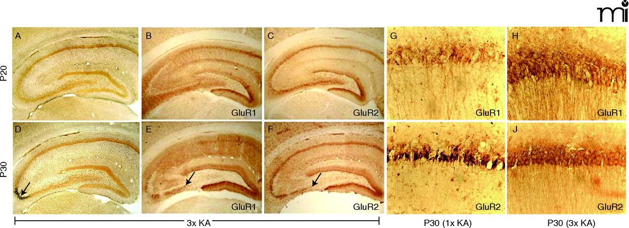
Figure 3.
Preservation of CA1 neurons and CA1 AMPA protein after 3×KA. A. Control photomicrographs of P20 silver stained section showing no argyrophilia. At P20 (A) and P30 (D) after 3×KA treatment, the CA1 was protected but CA3 damage shown as black staining was present. At P20 and P30, GluR1A (B, E) and GluR2B (C, F) protein levels were similar to controls (see Fig. 1⇑), except for in the CA3 region associated with cell loss (arrow). At P30, GluR1A immunolabeling (G) was significantly decreased in CA1 whereas GluR2B was increased (I). Steady levels of GluR1A and GluR2B were observed after 3×KA (H, J). Modified from (32, 45). Reprinted with permission.

