- Institution: Stanford Univ Med Ctr Lane Med Lib/Periodical Dept/Rm L109
- Sign In as Member / Individual
Revealing the Potential of Intranasally Administered Orexin A (Hypocretin-1)
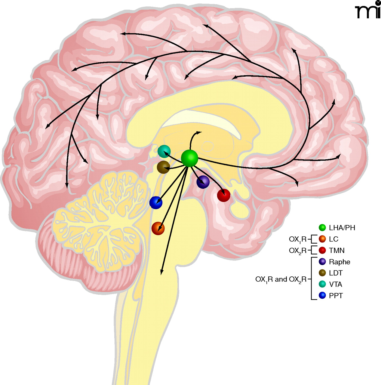
Main projections of orexin neurons. This figure summarizes orexinergic projections in the human brain as predicted from rodent studies. Circles show regions with strong expression of orexin receptors type 1 and type 2 (OX1R and OX2R, respectively) and dense orexinergic projections. Orexin neurons originating in the lateral hypothalamic area (LHA) and posterior hypothalamus (PH) regulate sleep and wakefulness and the maintenance of arousal by sending excitatory projections to the entire CNS, excluding the cerebellum, with particularly dense projections to monoaminergic and cholinergic nuclei in the brain stem and hypothalamic regions, including the locus coeruleus (LC, containing noradrenaline), tuberomammillary nucleus (TMN, containing histamine), raphe nuclei (Raphe, containing serotonin) and laterodorsal/pedunclopontine tegmental nuclei (LDT/PPT), containing acetylcholine). Orexin neurons also have links with the reward system through the ventral tegmental area (VTA, containing dopamine) and with the hypothalamic nuclei that stimulate feeding behavior. Figure from (1). Used with permission.

