|
 
 |
| ORIGINAL ARTICLE |
|
| Year : 2015 | Volume
: 3
| Issue : 1 | Page : 16-22 |
|
Effect of desensitizers on retention of castings cemented to anatomically prepared teeth
Manish R Chauhan, Arti P Wadkar
Department of Prosthodontics and Crown and Bridge, Nair Hospital Dental College, Mumbai, Maharashtra, India
| Date of Web Publication | 5-Jan-2015 |
Correspondence Address:
Dr. Manish R Chauhan
A/304, Aabhas Complex, Behind Ramdevpeer Temple, Near Garden View Hospital, Tulinj Road, Nallasopara (East), Thane, Mumbai - 401 209
India
 Source of Support: None, Conflict of Interest: None  | Check |
DOI: 10.4103/2347-4610.148515

Context: The extensive use of desensitizing agents to control incidences of post-cementation sensitivity can variably affect the forces required for debonding of the prosthesis. The available literature offers inconsistent results and is generally carried out using a flat occlusal preparation. Aims: Evaluation of effect of desensitizing agents-nonpolymerizable (Systemp) and polymerizable (Clearfil SE Bond)-on retention of complete veneer castings luted to anatomically prepared teeth with zinc phosphate and glass ionomer cement (ZPC and GIC). Materials and Methods: Seventy maxillary second premolars were prepared conforming to the anatomic form to receive complete veneer castings. Two sets of dies were prepared; one for obtaining castings and second to permit mathematical calculation of the surface area. The specimens were divided into four experimental groups of 15 samples each and two control groups of 5 samples each. Two desensitizers and two luting cements were used. After cementation, the samples were tested at crosshead speed of 0.5 mm/min on universal testing machine (UTM) using a customized self-aligning device. Minimum load required to debond the specimens was recorded. Force value for each specimen was divided by its surface area to obtain the tensile bond strength. Results: Significant reduction in tensile bond strength was noted for all the experimental groups, irrespective of luting cement used. Conclusion: Systemp-glass ionomer group exhibited highest bond strength followed by Systemp-zinc phosphate, Clearfil SE Bond-glass ionomer and Clearfil SE Bond-zinc phosphate group. Keywords: Dentin hypersensitivity, desensitizing agent, tensile bond strength
How to cite this article:
Chauhan MR, Wadkar AP. Effect of desensitizers on retention of castings cemented to anatomically prepared teeth. Eur J Prosthodont 2015;3:16-22 |
| Introduction | |  |
Dentin hypersensitivity is caused by exposure of dentin commonly resulting from either removal of enamel or denudation of the root surface by loss of cementum and overlying periodontal tissues. This may occur due to various reasons such as root planing, cavity preparation, or tooth preparation for receiving crowns. Dentin hypersensitivity after cementation of interim restorations has been attributed to microleakage and formation of bacterial by-products. However, hypersensitivity has been reported even after cementation of definitive prostheses with the most widely used luting agents like zinc phosphate and glass ionomer cement (ZPC and GIC). [1],[2] Hence, there has been an extensive use of polymerizable and nonpolymerizable desensitizing agents to control the same.
The polymerizable products like dentin bonding agents seal the orifices of the exposed tubules; whereas, the nonpolymerizable formulations coat the surface of the dentinal tubules and prevent fluid movement, thereby reducing sensitivity. However, their effect on the properties of the luting cements has not been studied extensively. Although retention of prosthesis is mainly determined by the geometric form of prepared tooth, the interplay between mechanism of action of desensitizer and mode of attachment of the luting agent can have variable effect on the resultant forces responsible for debonding of the cemented castings. The null hypothesis for the present study was that polymerizable and nonpolymerizable desensitizers do not have any effect on the force responsible for dislodging complete veneer base-metal ceramometal castings luted with ZPC and GIC.
| Materials and Methods | |  |
Selection and mounting of specimens, tooth preparation, fabrication of wax patterns and metal copings, cementation, storage conditions and the tensile testing conditions were standardized to minimize the effect of variable factors on the observations and the final result.
Seventy human maxillary second premolars extracted for orthodontic purpose with minimum crown height of 6.5 mm were collected. The specimens were divided into four experimental groups and two control groups as shown in [Table 1].
Each tooth was vertically embedded in self-cure acrylic resin with a preformed metal mold used as a matrix. Tooth preparation conforming to the anatomic form and contour as against a flat occlusal surface used by previous investigators [3],[4],[5],[6],[7],[8] was done using two separate aluminium attaching jigs which permit the handpiece to be held in various fixed positions [Figure 1]. The occlusal surface was prepared conforming to the anatomic form in such a way that the central groove was 1 mm below the cusp tips. The total occlusal convergence was maintained at 4° as measured with Nikon measuroscope, Japan [Figure 2]. Goodacre et al., recommended a minimum finish line depth of approximately 0.3 mm for all metal crowns. [9] Therefore, a 0.5 mm shoulder gingival finish line was prepared above the cementoenamel junction (CEJ).
Thus, all the prepared specimens had a constant taper and height, but with variable resultant size of individual tooth [Figure 3]. The prepared teeth were approximately elliptical in cross-section [Figure 4] and without a functional bevel so as to have geometric symmetry and permit mathematical calculation of the surface area.
Impressions were made in addition silicone (Elite HD +; Zhermack, Italy) and two sets of dies were prepared. To provide a connection for the universal testing machine (UTM), 0.5 mm thick copings with round loop were made [Figure 5]. Castings were checked for good fit on the prepared crowns. Systemp desensitizer was applied on Group I specimens by brushing onto the tooth structure for 10 s on each surface using a disposable microbrush (Microbrush International, USA). Clearfil SE Bond (Kuraray Medical Inc), a sixth generation bonding agent with one bottle containing the self-etch primer and the other containing the bond was applied on Group II specimens. The primer was first applied on the tooth surface using a microbrush and maintained for 20 s. The bond was then applied and light-cured for 10 s using light-emitting diode (LED) light-curing unit, Gnatus on every surface. Group I-A and Group II-A specimens were cemented with ZPC and Group I-B and Group II-B specimens were cemented with GIC under a static load of 5 kg, maintained for about 7 min, corresponding to the setting time of the cement.
A customized self-aligning device consisting of a flexible wire rope welded to solid metal spheres at both the ends [Figure 6] was used to permit alignment of the specimen along the apicocoronal axis of the tooth, as opposed to rigid attachments used by past workers. [4],[7],[10] A center screw made in brass served as the mode of attachment to the Hounsfield UTM, which in turn was connected to the force transducer. The samples were tested at a crosshead speed of 0.5 mm/min until failure. [11],[12],[13] The minimum load required to debond the specimen was recorded in Newton.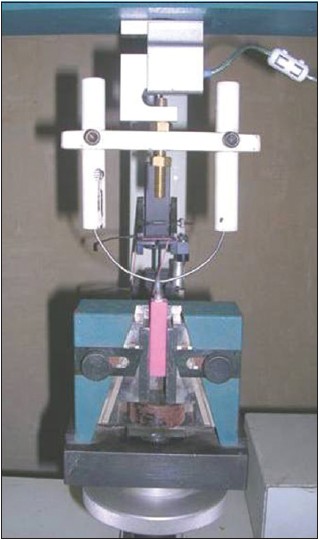 | Figure 6: Testing on UTM using self-aligning device. UTM = Universal testing machine
Click here to view |
Calculating the surface area of prepared teeth
The prepared maxillary premolar is not a regular geometrical figure, but closely resembles a solid truncated cone with an elliptical cross-section. Kaufman et al., [14] and Shillingburg [15] from their studies concluded that the retentive force increases with increase in the surface area.The total surface area was calculated mathematically by addition of the lateral surface area and the occlusal surface area.
The lateral surface area (S) of an elliptical cone is given by the formula

Where 'a' is the semi-major axis, 'b' is the semi-minor axis, 'h' is the height of the cone, and E is given by the formula

Since we are measuring the surface area of a solid complete cone, the angle 'v' is 360°.
Hence, cos 2v = cos 2 360° =1 and sin 2v = sin 2 360° =0

The surface area of each prepared tooth was calculated [Figure 7].
Step 1.
Calculating the lateral surface area of the larger cone 'S' and smaller cone 'S 0 '
Step 2.
Determining the lateral surface area of the tooth 'S − S 0 '
Step 3.
Deducting the area of two triangles observed on the proximal view of the tooth
Step 4.
Adding the surface area of the occlusal surface resembling a geometric ellipse

where p and p 0 are the semi-major axes of semi-ellipse E and semi-ellipse E 0 , respectively and q is the semi-minor axis common to both the ellipses.
| Results | |  |
[Table 2], [Table 3], [Table 4] show the values of force, surface area and the calculated tensile bond strength of specimens of the six groups.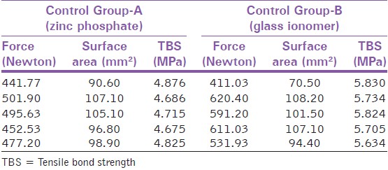 | Table 4: Comparison of tensile bond strength of control Group A and control Group B
Click here to view |
Statistical analysis using unpaired t-test was carried out [Table 5].
On the basis of observations, the following results were obtained:
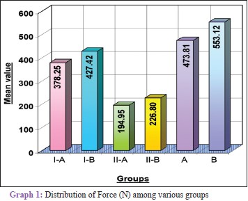
- Graph 1 shows that the dislodging force was dependent on the desensitizing agent applied and the luting cement used. Therefore, there was a marked variation in intergroup values
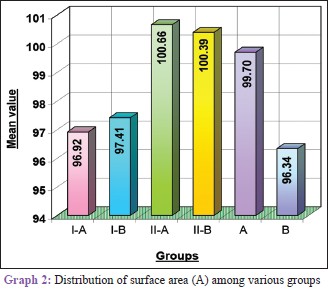
- Graph 2 indicates the difference in the mean surface area between the various groups. It was not statistically significant
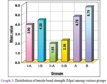
- Graph 3 indicates that the mean tensile bond strength values for all experimental groups were significantly less than those obtained for control groups. The results indicated that the tensile bond strength was reduced irrespective of the desensitizer and the luting cement used, but to a varying degree.
| Discussion | |  |
The retentive strength of cemented crowns depends on the physicochemical properties of the luting agent used. The desensitizing agents applied on the tooth surface can however, alter the retentive properties of the cementing medium, thereby compromising the restoration. Hence, this study was undertaken to determine the effect of two groups of desensitizing agents-nonpolymerizable (Systemp desensitizer) and polymerizable (Clearfil SE Bond)-on the retention of complete veneer base-metal ceramometal castings luted with commonly used luting agents-ZPC and GIC.
Ayad et al., [16] used a milling machine for standardizing axial tooth reduction; whereas, the occlusal surface was made flat. They reported mean force values of 213.9 and 308.5 N for ZPC and GIC, respectively. These values were significantly lower than the values obtained in the present study, which were 473.81 and 553.12 N, respectively for the two control groups. This could be attributed to the lesser height of 3.5 mm and the flat occlusal surface prepared by them, as compared to 4 mm axial height and anatomic preparation of occlusal surface in the present study.
Tuntiprawon [4] used a similar milling machine to prepare teeth with 3 mm height, 6° convergence angle, and a flat occlusal surface. The specimens were tensile tested using Lloyd TM with a rigid attachment device. He reported a mean value of 343.98 N for ZPC and 482.04 N for GIC in experimental Group II. These were considerably lower than the values obtained in the present study. This could be explained on the basis of lesser preparation height of 3 mm and a flat occlusal surface prepared by him. The use of rigid attachment for testing procedures could have had a role in the lower values displayed.
The prepared teeth were not acid-etched prior to cementation of castings. It has been reported by Stewardson et al., [17] that Systemp desensitizer was effective in reducing pain from hypersensitivity and this finding was unaffected whether or not the tooth was acid-etched prior to the application of the agent. The second desensitizer used in the study was Clearfil SE Bond which contains a self-etching primer. Hence, separate etching was not done in any of the groups.
Yim et al., [18] employed controlled crown preparation surface area and evaluated the effect of All-Bond 2 and Gluma desensitizer on retention of full crowns using standardized preparations when cemented with zinc phosphate, glass ionomer, resin-modified glass ionomer, and resin cements. They maintained a flat occlusal surface with 26° total occlusal convergence and 4 mm axial height. They reported mean tensile bond strength values of 1.68 ± 0.08 and 2.36 ± 0.20 MPa with ZPC and GIC, respectively. These were significantly lower than the values obtained in this study (3.9 and 4.39 MPa, respectively), which could be the result of flat occlusal surface and preparation convergence angle of 26°. They concluded that Gluma desensitizer significantly reduces crown retention with both ZPC (0.81 ± 0.11 Mpa) and GIC (1.98 ± 0.23 MPa). All-Bond 2 resulted in the lowest bond strength for crowns cemented with zinc phosphate (0.67 ± 0.14 MPa). The decrease in bond strength in all three groups was in correlation with the findings of the present study.
Sharma et al., [13] reported a decrease in retentive values when a resin-based sealer (One Step) was used with ZPC and an increase in retention with GIC. Johnson et al., [19] also concluded that resin sealer (One Step) decreased the casting retentive stress by 42% when used with zinc phosphate. This was slightly lesser than the results obtained in this study, which was approximately 59.45%. However, their finding regarding 55% increase in retention when sealer was used in conjunction with GIC, was inconsistent with the results of this study.
The mean retentive value for castings cemented with zinc phosphate (control A) was 4.76 ± 0.09 MPa. This was consistent with the values obtained by Zidan and Ferguson [11] who conducted experiment on teeth with a height of 4 mm and 6° taper concurrent with the present study. These values were higher when compared to results obtained by Gorodovsky and Zidan [5] (3.08 ± 0.9 MPa), Johnson et al., [6] (3.7 ± 1.0 MPa), and Yim et al., [18] (1.68 ± 0.08 MPa). This could be attributed to the decreased taper of 4-5° followed in the present study as compared to that used by Yim et al., [18] (26°) and Johnson et al., [6] (20°). All the above authors had prepared a flat occlusal surface. It can therefore be concluded that the anatomic tooth reduction has a direct correlation with the higher bond strength values exhibited in this study.
The mean retentive value for castings cemented with GIC in this study (5.75 ± 0.08 MPa) was significantly higher than results obtained in the previous studies by Zidan and Ferguson [11] (4.72 MPa), Gorodovsky and Zidan [5] (3.12 ± 1.2), Johnson et al., [6] (2.7 ± 1.2), and Yim et al., [18] (2.36 ± 0.20). The common finding in their methodology was a flat occlusal surface. Hence, it can be inferred that a flat occlusal surface can potentially decrease the bond strength values. The smaller values obtained by Johnson et al., [6] and Yim et al., [18] as compared to other authors could also be due to the increased amount of taper (20 and 26°, respectively) used by them.
There was a reduction of about 18.06% with Systemp and 59.45% reduction with Clearfil SE Bond desensitizer used in conjunction with ZPC. The decrease in the mean stress values was in correlation with the results obtained by Mausner et al., [10] Yim et al., [18] and Johnson et al. [19]
The results showed a decrease of about 23.66% with Systemp and 61.7% with Clearfil SE Bond used with GIC. The decrease in values was consistent with previous studies performed by Mausner et al., [10] and Yim et al. [18] Sipahi et al., studied the effects of precementation desensitizing laser treatment and conventional desensitizing agents on crown retention. They reported a 38% decrease in retention when Gluma desensitizer was used in conjunction with GIC as compared to control group. [20]
The results were inconsistent with the findings of Swift et al., [7] who studied the effect of resin primers and adhesives on the retention of crowns. They concluded that the two agents used in the study, that, Gluma desensitizer or One Step had little or no effect on the retention of crowns luted with zinc phosphate, glass ionomer, or resin-modified GIC. The results were also inconsistent with the findings of Patil et al., [21] who reported no effect of Gluma desensitizer on retention with ZPC or GIC.
Similarly, the results did not conform to the findings of Johnson et al., [6] who studied the effect of a non-resin sealer glutaraldehyde-based desensitizer (Gluma) on the retention of cemented castings and concluded that there was no effect on crown retention for zinc phosphate, glass ionomer, and resin-modified cement. The results were contradictory to the study by Sonune et al., [12] who reported an increase in retentive values when Gluma desensitizer was used with ZPC and GIC.
After relating all the data inferred, the results of this study indicate that the tensile bond strength values in the experimental groups were much less than the corresponding control groups, indicating a loss of retention irrespective of the type of desensitizer and the luting cement used. Therefore, the use of desensitizing agents should be restricted in clinical practice.
Zinc phosphate attains its retentive qualities by filling irregularities in the prepared dentin and internal surface of the casting. The desensitizing agents form a smooth coat on the tooth surface which might interfere with mechanical interlocking of the luting cement because of decrease in number of cement locks.
GIC has an adhesive bond to the tooth structure, the polyacrylic acid reacting with the calcium ions of the hydroxyapatite. When polymeric resins are used as desensitizer, they seal the tubules and interact with the altered intertubular dentin. GIC cannot then optimally react with the dentin.
One of the prominent findings of this study was the increase in bond strength values when the occlusal surface was prepared according to the biomechanical principles, conforming to the anatomic form as against a flat surface. It is therefore recommended that further research work be conducted with reproduction of anatomic form which can provide more comprehensive results, with better clinical acceptance.
The study was conducted in vitro trying to simulate the oral conditions. However, the effects of thermocycling and cyclic loading could not be evaluated. Although all efforts were made to standardize the tooth preparation procedure to allow a near accurate mathematical calculation of the surface area, there are methods employing electron microscope attached to computer software which can eliminate the negligible amount of error and give more accurate values. However, the use of these sophisticated instruments was not within the purview of this study. It is also proposed that further studies be conducted with different combinations of desensitizers and luting cements on a larger sample size so as to obtain more authentic results which can find greater acceptance.
| Conclusion | |  |
Within the limitations of this study, it can be concluded that:
- The use of desensitizers should be restricted, since the bond strength decreased with both Systemp and Clearfil SE Bond when used in conjunction with either zinc phosphate or GIC
- GIC should be preferred over zinc phosphate cement whenever used with a desensitizer
- Anatomic tooth preparation must be followed since the bond strength values were significantly higher when a tooth was prepared anatomically as compared to a flat occlusal surface.
| References | |  |
| 1. | Kern M, Kleimeier B, Schaller H, Strub JR. Clinical comparison of postoperative sensitivity for a glass ionomer and a zinc phosphate luting cement. J Prosthet Dent 1996;75:159-62.  |
| 2. | Bebermeyer RD, Berg JH. Comparison of patient-perceived postcementation sensitivity with glass ionomer and zinc phosphate cements. Quintessence Int 1994;25:209-14.  |
| 3. | el-Mowafy OM, Fenton AH, Forrester N, Milenkovic M. Retention of metal ceramic crowns cemented with resin cements: Effects of preparation taper and height. J Prosthet Dent 1996;76:524-9.  |
| 4. | Tuntiprawon M. Effect of tooth surface roughness on marginal seating and retention of complete metal crowns. J Prosthet Dent 1999;81:142-7.  |
| 5. | Gorodovsky S, Zidan O. Retentive strength, disintegration, and marginal quality of luting cements. J Prosthet Dent 1992;68:269-74.  |
| 6. | Johnson GH, Lepe X, Bales DJ. Crown retention with use of a 5% glutaraldehyde sealer on prepared dentin. J Prosthet Dent 1998;79:671-6.  |
| 7. | Swift EJ Jr, Lloyd AH, Felton DA. The effect of resin desensitizing agents on crown retention. J Am Dent Assoc 1997;128:195-200.  |
| 8. | Tay FR, Pashley DH, Mak YF, Carvalho RM, Lai SC, Suh BI. Integrating oxalate desensitizers with total-etch two-step adhesive. J Dent Res 2003;82:703-7.  |
| 9. | Goodacre CJ, Campagni WV, Aquilino SA. Tooth preparations for complete crowns: An art form based on scientific principles. J Prosthet Dent 2001;85:363-76.  |
| 10. | Mausner IK, Goldstein GR, Georgescu M. Effect of two dentinal desensitizing agents on retention of complete cast coping using four cements. J Prosthet Dent 1996;75:129-34.  |
| 11. | Zidan O, Ferguson GC. The retention of complete crowns prepared with three different tapers and luted with four different cements. J Prosthet Dent 2003;89:565-71.  |
| 12. | Jalandar SS, Pandharinath DS, Arun K, Smita V. Comparison of effect of desensitizing agents on the retention of crowns cemented with luting agents: An in vitro study. J Adv Prosthodont 2012;4:127-33.  |
| 13. | Sharma S, Patel JR, Sethuraman R, Singh S, Wazir ND, Singh H. A comparative evaluation of the effect of resin based sealers on retention of crown cemented with three types of cement -an in vitro study. J Clin Diagn Res 2014;8:251-5.  |
| 14. | Kaufman EG, Coelho DH, Colin L. Factors influencing the retention of cemented gold castings. J Prosthet Dent 1961;11:487-502.  |
| 15. | Shillingburg HT. Fundamentals of fixed prosthodontics. 3 rd ed. Quintessence Publishing Co, Inc; 1997. p. 120.  |
| 16. | Ayad MF, Rosenstiel SF, Salama M. Influence of tooth surface roughness and type of cement on retention of complete cast crowns. J Prosthet Dent 1997;77:116-21.  |
| 17. | Stewardson DA, Crisp RJ, Mchugh S, Lendenmann U, Burke FJ. The effectiveness of Systemp desensitizer in the treatment of dentine hypersensitivity. Prim Dent care 2004;11:71-6.  |
| 18. | Yim NH, Rueggeberg FA, Caughman WF, Gardner FM, Pashley DH. Effect of dentin desensitizers and cementing agents on retention of full crowns using standardized crown preparations. J Prosthet Dent 2000;83:459-65.  |
| 19. | Johnson GH, Hazelton LR, Bales DJ, Lepe X. The effect of a resin-based sealer on crown retention for three types of cement. J Prosthet Dent 2004;91:428-35.  |
| 20. | Sipahi C, Cehreli M, Ozen J, Dalkiz M. Effects of precementation desensitizing laser treatment and conventional desensitizing agents on crown retention. Int J Prosthodont 2007;20:289-92.  |
| 21. | Patil PG, Parkhedkar RD, Patil SP, Bhowmik HS. Comparative evaluation of effect of polymerizable and non-polymerizable desensitizing agents on crown-retentive-strength of zinc-phosphate, glass-ionomer and compomer cements. Eur J Prosthodont Restor Dent. 2012;20:102-10.  |
[Figure 1], [Figure 2], [Figure 3], [Figure 4], [Figure 5], [Figure 6], [Figure 7]
[Table 1], [Table 2], [Table 3], [Table 4], [Table 5]
|