|
 
 |
|
REVIEW ARTICLE |
|
|
|
| Year : 2011 | Volume
: 17
| Issue : 1 | Page : 3-6 |
| |
Proteomics in obstetrics and gynecology
Seema Lekhwani1, Vijay Shankar1, ND Vaswani2
1 Department of Biochemistry, Pt. B. D. Sharma PGIMS, Rohtak, Haryana, India
2 Department of Pediatrics, Pt. B. D. Sharma PGIMS, Rohtak, Haryana, India
| Date of Web Publication | 18-Jun-2011 |
Correspondence Address:
Seema Lekhwani
9J/55, Medical Enclave, Pt. B. D. Sharma PGIMS, Rohtak - 124 001, Haryana
India
 Source of Support: None, Conflict of Interest: None
DOI: 10.4103/0971-6866.82185

 Abstract Abstract | | |
Proteomics helps to understand the basic biological processes critical to normal cellular functions as well as the development of diseases. It identifies the essential components of these processes and exploits these components as targets in the development of new methods to prevent or treat diseases. Proteomics, although in an infancy stage in India, has the potential to complement and further enlarge the wealth of information in medicine, especially in the field of cancer. This article reviews the recent progress in proteomic techniques and their applications in the field of obstetrics and gynecology.
Keywords: Biomarkers, gynecologic cancers, pre-eclampsia, prematurity, proteomic tools
How to cite this article:
Lekhwani S, Shankar V, Vaswani N D. Proteomics in obstetrics and gynecology. Indian J Hum Genet 2011;17:3-6 |
| Introduction | |  |
The Human Genome Project had made the scientific community and public believe that all biology and medicine would become understandable after the sequencing of the DNA. As biology is much more complicated than the structure and function of nucleic acids, the practical applications are quite limited. The study of living systems has now expanded past genomics based on the rationale that it is the protein products of the gene and not simply gene expression that have effects and cause disturbances at the cellular level. Its advent has provided the hope of discovering novel biological markers for use in the screening, early diagnosis and prediction of response to therapy. [1]
Mass spectrometers (MSs) like matrix-assisted laser desorption/ionization time of flight (MALDI-TOF) MS, surface-enhanced laser desorption/ionization time of flight (SELDI-TOF) MS, electro spray ionization MS and Fourier transform ion cyclotron resonance MS have enabled the study of molecular mechanisms of cancer with complex fluids and tissue samples with a good sensitivity and resolution. Proteomic technologies like two-dimensional (2D) polyacrylamide gel electrophoresis (PAGE), isotope-coded affinity tags, SELDI-TOF, multidimensional protein identification technology, protein arrays and protein chips promise application at the bedside for discovering protein patterns that distinguish disease from disease-free states with a high sensitivity and specificity. [2]
| Proteomics in Gynecology | | |
Cervical cancer is one of the leading causes of cancer morbidity and mortality in women worldwide. More than 98% of the cases are related to a Human papilloma virus (HPV) infection. Researchers are working on the identification and functional verification of host proteins associated with HPV E6 and E7 oncoproteins. This may provide useful information for understanding cervical carcinogenesis and development of cancer-specific markers. In addition, proteomic profiling of altered proteins by anticancer drugs on cervical cancer may contribute to providing the fundamental resources for investigation of disease-specific target proteins, elucidation of novel mechanisms of action and development of new drugs. [3]
To date, the quest to develop a non-invasive diagnostic test for endometriosis has mostly concentrated on the levels of cytokines and growth factors that are involved in inflammation, angiogenesis and tissue remodeling present in serum, peritoneal fluid, endometrium and endometriotic lesions. As this has not yet translated into the development of such a diagnostic test, proteomic techniques are now being employed to identify proteins that are potential biomarkers of the disease. [4]
The urgent need of biomarkers accurately detecting early-stage epithelial ovarian cancer has prompted many research groups to enter in a search for specific peptide signatures that may discriminate transformed cells from those of the normal ovarian microenvironment. CA-125, the most widely used biomarker for ovarian cancer, does not have a high positive predictive value, and it is only effective when used in combination with other diagnostic tests [Table 1]. [5] A combination of mass spectra generated by new proteomic techniques such as SELDI-TOF- and artificial intelligence-based informatic algorithms have been used to discover a small set of key proteins in order to discriminate between normal and patients with ovarian cancer. Serum protein pattern analysis might be applied ultimately in medical screening clinics as a supplement to thediagnostic work up and evaluation. [6],[7],[8],[9] Breast cancer is one of the most frequent and deadly cancers worldwide. Although survival of patients has increased over the last decade in relation to screening programs and post-operative adjuvant therapies (hormone therapy, chemotherapy), many patients die from metastatic relapse. Research studies of molecular alterations have successfully elucidated mechanisms of mammary oncogenesis and identified key genes such as ERBP-2, TPS3, CCND1, BRCA1 and BRCA2. It has now allowed considerable therapeutic progress by targeting hormone receptors as well as ERBB2 and HER2 receptors. [10] A proteomic-based approach utilizing 2D-PAGE and newer technologies such as laser capture microdissection and highly sensitive mass spectrometry methods are currently being used together to identify lower abundance proteins that are differentially expressed between defined cell populations. High-density protein arrays, antibody arrays and lysate arrays are novel technologies still in development phases, which will enable identification of validated targets in small biopsy specimens. SELDI-TOF analysis enables the high-throughput characterization of lysates from few tumor cells, and may be best suited for clinical biomarker studies. [11] Breast cancer studies have used tumor tissue and also biological fluids including serum, plasma, nipple aspirate or ductal lavage material. Breast cancer proteomics has already identified the markers of potential clinical interest (such as the molecular chaperone 14-3-3 sigma). With examples of fibroblast growth factor and H19, an oncogenic, non-coding mRNA, proteomics have become a powerful approach for deciphering the complex signaling circuitry involved in tumor growth. [12],[13] Rui et al. found HSP27 upregulated while 14-3-3 sigma downregulated in serum of breast cancer patients. These two biomarkers can also be used for further study in drug design and breast cancer therapy. [14] Recently, Liu et al. supported that the analysis of Ca ++ binding protein S100A11 expression in breast cancer may be an effective tool that helps in the detection of early-stage breast cancer, and it is a novel diagnostic marker in breast carcinoma. [15]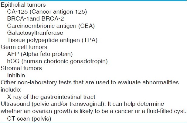 | Table1: Diagnostic tests of symptomatic women shown to be positive in with ovarian cancer
Click here to view |
| Proteomics in Obstetrics | | |
Pre-eclampsia is a pregnancy-specific syndrome and a major cause of maternal mortality. The pathophysiology of pre-eclampsia is unknown and no proteome analysis is reported. Watanabe et al. sought to identify proteins associated with pre-eclampsia using 2D electrophoresis on sera of six patients with pre-eclampsia and six normal pregnant women, followed by comparison of the profiles. Clusterin was identified by MALDI-TOF-MS followed by peptide mass fingerprinting, a protein database search and Western blot analysis. [16] Mine and colleagues found dynactin, a protein related to cell turnover in placental proteome maps, by novel 2D-immunoblotting analysis. [17] Myers et al. carried out a pilot study to use protein chip technology to determine differences in protein profiles in plasma taken at 26 weeks from women at risk of developing pre-eclampsia. They found five proteins upregulated significantly in samples from women who subsequently developed pre-eclampsia compared with women who remained normotensive. [18] Recent evidence suggests that a major cause of prematurity-associated neonatal pathology is the fetal and neonatal response to inflammation or infection rather than respiratory distress syndrome, intraventricular hemorrhage, necrotizing enterocolitis and bronchopulmonary dysplasia as the primary causes of pre-term delivery. Proteomic profiling of amniotic fluid provides a precise means for detection of inflammation by revealing the presence of four biomarkers (defensins-2 and 1, Calgranulin C and A) that are highly predictive of intrauterine inflammation (MR score). MR score presents a gradient of disease activity progressing from "absent," "mild" to "severe" inflammation. Thus, it provides the ability to identify patients who might benefit from interventions in utero in a modern diagnostic-therapeutic framework. [19]
Others
Proteomic tools are being applied in varied fields related with reproductive function, such as the study of oocyte maturation, spermatogenesis and fertilization in mammals. [20],[21],[22],[23] The identification of specific genes in oocytes and embryos is now possible with the use of powerful tools such as library analysis or subtractions, DNA array, comparative analysis of databanks from other mammals and 2D-gel electrophoresis analysis. Finally, RNA interference is a useful tool for studying gene function by knocking out the activity of specific genes, and will be used in oocytes and embryos. [24] Amniotic fluid is a potential source of biomarkers for many disorders that may occur during pregnancy or for embryonic abnormalities. Proteomics have already been applied in the analysis of tissues from fetuses with Down's syndrome. [25] The proteomic analysis of follicular fluid by Angelucci et al. revealed identification of a large number of acute phase proteins, including transferrin, ceruloplasmin, afamin, hemopexin, haptoglobin and plasma amyloid protein, suggesting the hypothesis that mammalian ovulation can be compared with an inflammatory event. Several important antioxidant enzymes, i.e. catalase, superoxide dismutase, glutathione transferase, peroxisonase, heat shock protein 27 and protein disulfide isomerase, were also identified. This indicates that, during maturation, the human follicle is well protected against toxic injury due to oxidative stress. [26]
| Conclusion | | |
Looking back, it seems that the major outcome of human genome sequencing has finally been to open the way to exploration of the proteome. Perhaps the most promising outcome of the work reported by scientists is the interface between proteomic technology and bioinformatics. The rapid explosion in the amount of data being generated by current genomic and proteomic technology already exceeds the analytical capacity of the human mind. The need for increasing sophistication in data management and statistical interpretation and attention to this aspect of research is critical to its successful translation into clinical practice. [27]
| References | | |
| 1. | Hondermarck H. Breast cancer: When proteomics challenges biological complexity. Mol Cell Proteomics 2003;2:281-91. 
|
| 2. | Somiari RI, Somiari S, Russell S, Shriver CD. Proteomics of breast carcinoma. J Chromatogr B Analyt Technol Biomed Life Sci 2005;815:215-25.
|
| 3. | Yim EK, Park JS. Role of proteomics in translational research in cervical cancer. Expert Rev Proteomics 2006;3:21-36.
|
| 4. | Poliness AE, Healy MG, Brennecke SP, Moses EK. Proteomic approaches in endometriosis research. Proteomics 2004;4:1897-1902.
|
| 5. | www.labtestsonline.org/understanding/conditions/ovarian-2.html
|
| 6. | Jones MB, Krutzsch H, Shu H, ZhaoY, Liotta LA, Kohn EC, et al. Proteomic analysis and identification of new biomarkers and therapeutic target for invasive ovarian cancer. Proteomics 2002;2:76-84.
|
| 7. | Ardekani AM, Liotta LA, PetricoinIII EF. Clinical potential of proteomics in the diagnosis of ovarian cancer. Expert Rev Mol Diagn 2002;2:312-20.
|
| 8. | Jacobs IJ, Menon U. Progress and challenges in screening for early detection of ovarian cancer. Mol Cell Proteomics 2004;3:355-66.
|
| 9. | Tchabo NE, Guancial EA, Czechowicz JA, Kohn EC. The role of proteomics in the diagnosis and treatment of ovarian cancer. Women′s Health (Lond Engl) 2005;1:365-74.
|
| 10. | Bertucci F, Birnbaum D, Goncalves A. Proteomics of breast cancer-principles and potential clinical applications. Mol Cell Proteomics 2006;5:1772-86.
|
| 11. | Wulfkuhle JD, McLean KC, Paweletz CP, Sgroi DC, Trockj BJ, Steeg PS, et al. New approaches to proteomic analysis of breast cancer. Proteomics 2001;1:1205-15.
|
| 12. | Hondermarck H, Vercoutter-Edouart AS, Revillion F, el-Yazidi-Belkoura I, Nurcombe V, Peyart JP. Proteomics of breast cancer for marker discovery and signal pathway profiling. Proteomics 2001;1:1216-32.
|
| 13. | Yazidi-Belkoura I, Adriaenssens E, Vercoutter-Edouart AS, Lemoinie J, Nurcombe V, Hondermarck H. Proteomics of breast cancer: Outcomes and prospects. Technol Cancer Res Treat 2002;1:287-96.
|
| 14. | Rui Z, Jian-Guo J, Yuan-Peng T, Hai P, Bing-Gen R. Use of serological proteomic methods to find biomarkers associated with breast cancer. Proteomics 2003;3:433-9.
|
| 15. | Liu XG, Wang XP, Li WF, Yang S, Zhou X, Li SJ, et al. Ca2+-binding protein S100A11: A novel diagnostic marker for breast carcinoma. Oncol Rep 2010;23:1301-8.
|
| 16. | Watanabe H, Hamada H, Yamada N, Sohda S, Yamakawa-Kobayashi K, Yoshikawa H, et al. Proteomic analysis reveals elevated serum levels of clusterin in patients with Preeclampsia. Proteomics 2004;4:537-43.
|
| 17. | Mine K, Katayama A, Nishino T, Kuwabara Y, Ishikawa G, Murata T, et al. Proteomic analysis of human placentae: Pre-eclampsia versus normal pregnancy. Placenta 2007;28:676-87.
|
| 18. | Myers J, Macleod M, Zreed B, Harris N, Mires G, Baker P. Use of proteomic patterns patterns as a novel screening tool in pre-eclampsia. J Obstet Gynaecol 2004;24:873-4.
|
| 19. | Buhimschi CS, Weiner CP, Buhimschi IA. Clinical proteomics: A novel diagnostic tool biology of preterm labor. Obstet Gynecol Surv 2006;61:481-6.
|
| 20. | Coonrod SA, Calvert ME, Reddi PP, Kasper EN, Digilo LC, Herr JC. Oocyte proteomics: Localization of mouse zona pellucida protein 3 to the plasma membrane of ovulated mouse eggs. Reprod Fertil Dev 2004;16:69-78.
|
| 21. | Yurttas P, Morency E, Coonrod SA. Use of proteomics to identify highly abundant maternal factors that drive the egg-to-embryo transition. Reproduction 2010;139:809-23.
|
| 22. | Sato k, Iwasaki T, Sakakibara K, Itakura S, Fukami Y. Towards the molecular dissection of fertilization signaling: Our functional genomic/proteomic strategies. Proteomics 2002;2:1079-89.
|
| 23. | Paza M, Morinb M, Mazo DJ. Proteome profile changes during mouse testis development. Comparative biochemistry and physiology Part D: Genomics and proteomics. 2006;1:404-15.
|
| 24. | Sirad MA, Dufort I, Coenen K, Tremblay K, Massicotte L, Robert C. The use of genomics and proteomics to understand oocyte and early embryo functions in farm animals. Reprod Suppl 2003;61:117-29.
|
| 25. | Tsangaris GT, Kolialexi A, Karamessinis PM, Anagnostopoloulos AK, Antsakalis A, Fountoulakis M, et al. The normal human amniotic fluid supernatant proteome. In Vivo 2006;20:479-90.
|
| 26. | Angrlucci S, Ciavardelli D, Giuseppe FD, Eleuterio E, Sulpizio M, Tiboni GM, et al. Proteome analysis of human follicular fluid. Biochim Biophys Acta 2006;1764:1775-85.27.
|
| 27. | Daly MB, Ozols RF. The search for predictive patterns in ovarian cancer: Proteomics meets bioinformatics. Cancer cell 2002;1:111-2.
|
[Table 1]
|


