|
 
 |
|
CASE REPORT |
|
|
|
| Year : 2012 | Volume
: 18
| Issue : 2 | Page : 256-258 |
| |
Unusual manifestation of Marden-walker syndrome
Amar M Taksande, KY Vilhekar
Department of Paediatrics, Jawaharlal Nehru Medical College, Datta Meghe Institute of Medical Sciences (DMIMS), Sawangi Meghe, Wardha, India
| Date of Web Publication | 8-Sep-2012 |
Correspondence Address:
Amar M Taksande
Department of Pediatrics, Jawaharlal Nehru Medical College, Sawangi (M), Wardha, Maharashtra-442 102
India
 Source of Support: None, Conflict of Interest: None
DOI: 10.4103/0971-6866.100798

 Abstract Abstract | | |
Marden-Walker syndrome (MWS) is characterized by multiple joint contractures, a mask-like face with blepharophimosis, micrognathia, high-arched or cleft palate, low-set ears, decreased muscular bulk, arachnodactyly, and kyphoscoliosis. We report a case of MWS along with unusual manifestation of neurological, cardiovascular, and genitourinary system.
Keywords: Arthrogryposis, atrial septal defect, inguinal hernia, joint contracture, Marden-Walker syndrome
How to cite this article:
Taksande AM, Vilhekar K Y. Unusual manifestation of Marden-walker syndrome. Indian J Hum Genet 2012;18:256-8 |
| Introduction | |  |
Arthrogryposis multiplex congenita (AMC) is diagnosed when two or more joints in more than one limb are fixed from birth. AMC may be caused by neurological and non-neurological causes. [1] Marden Walker Syndrome (MWS) is an autosomal recessive disorder characterized by multiple joint contractures, a mask-like face with blepharophimosis, micrognathia, high-arched or cleft palate, low-set ears, decreased muscular bulk, arachnodactyly, kyphoscoliosis, decreased muscular bulk, failure to thrive, and marked motor and mental retardation. [2] Here we report a typical case of MWS to highlight three interesting and unusual findings occurring in the same patient, viz., neurological manifestation, congenital heart disease, and genitourinary system.
| Case Report | | |
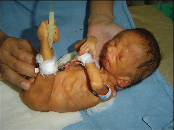
A 34-week preterm male baby was born to a 24-year-old second gravida mother by caesarean section of a non-consanguineous marriage. The antenatal period was uneventful. There was no history of exposure to any known teratogen. The antenatal ultrasonography examination (done at 28 weeks) was reportedly oligohydramnios. The baby did not suffer perinatal asphyxia. The child at birth weighed 1.5kg, head circumference was 34cm, and he was noted to have deformed limbs with flexion and extension joint contracture. Active and passive mobility of the joints was restricted. There was a limited motor function in the shoulder, elbow, and wrist joints. Bilateral internal rotation of the shoulder joint and flexion of the elbow joints was present. Arachnodactyly with bilateral wide gap between index finger and middle finger was noted. In lower limbs, bilateral hips were fixed in flexion, knees were extended, and ankles were dorsiflexed [Figure 1]. Congenital talipes equinovarus deformity was noted in the left foot. Facies deformities include asymmetric face, low-set ears, high-arched palate, short neck, and micrognathia was present. The muscles were markedly hypoplastic in the upper and lower limbs. Hypospadias with bilateral inguinal hernia was present. MRI head revealed dilatation of the ventricles suggestive of hydrocephalus with hypoplastic brainstem and hypoplasia of the inferior vermis and cerebellar hemispheres. Abdominal sonography was normal. Ocular ultrasonography was normal. Echocardiography revealed small-size ostium secundum atrial septal defect without pulmonary hypertension. Renal function tests and serum creatinine phosphokinase were normal. The karyotype was normal. No abnormality of any other internal organs was detected. A skin biopsy, EMG, and muscle biopsy, was declined by the father of the child. Specific inquiry regarding the presence of a similar disorder in the family or relatives yielded a negative result. On the basis of phenotypic features, a diagnosis of MWS was made.
| Discussion | | |
AMC is not a disease but refers to a group of birth defects characterized by multiple joint contractures. The syndrome is caused by neuropathic disease, myopathic disease, or any other cause of decreased fetal joint mobility. Neurological causes of AMC include: trisomy 13 and 18, Smith-Lemli-Opitz syndrome, Zellweger syndrome, MWS, spinal cord injury, amyoplasia congenita, infantile spinal muscular atrophy, infantile neuronal degeneration, focal infantile spinal muscular atrophy, Moebius syndrome, congenital hypomyelinating neuropathy, and craniocarpotarsal dysplasia. Non-neurological causes of AMC are cartilaginous abnormalities and physical constraint to movement. [1]
MWS was reported initially in 1966 by Marden and Walker, who described a female infant with blepharophimosis, micrognathia, immobile facies, kyphoscoliosis, limb contractures, pigeon breast, and arachnodactyly. [3] In 1991, Linder et al. recognized a case of MWS in the neonatal period. [4] To date, 30 MWS-affected individuals have been reported. [5] It is inherited as an autosomal recessive trait and most signs are present in the neonatal period. [6] The most frequent signs include multiple joint contractures, dysmorphic features with a mask-like face, blepharophimosis, ptosis, micrognathia, cleft or high arched palate, low-set ears, arachnodactyly, and decreased muscle mass. Camptodactyly, chest deformation as pectus (excavates or carinatus), kyphoscoliosis, and absent deep tendon reflexes are frequent. Minor malformations consist of renal anomalies [7] (renal cystic dysplasia, unilateral kidney hypoplasia, unilateral mild hydronephrosis), cardiovascular abnormalities (dextrocardia, ventricular septal defect), hypospadias, omphalomesenteric duct, hypertrophic pyloric stenosis, [8] duodenal bands, hypoplastic right lower lobe of the lung, displacement of the larynx to the right, and vertebral anomalies. Cerebral malformations such as hydrocephalus, hypolastic corpus callosum, cerebellar vermis hypoplasia, and enlarged cisternal magna maybe associated with microcephaly have also been described. [6]
The diagnosis of MWS is based on clinical criteria.Arthrogryposis can be picked up antenatally on ultrasound from the second trimester onwards. In pregnancy, oligohydramnios should indicate more detailed ultrasonographic examination, as ankylosed joints can be detected in utero. In 1995, Ben-Neriah et al. made the prenatal diagnosis of MWS in a fetus with a previously affected sibling by finding intrauterine growth retardation and renal cystic disease. [7] The disease course is always characterized by failure to thrive and psychomotor retardation. Mental retardation remains severe but contracture is not progressive and decreases with advancing age and physiotherapy. Death has occurred at approximately 3 months of age because of aspiration, sepsis, or cardiac failure. The treatment is only symptomatic with multidisciplinary management.
| References | | |
| 1. | Hageman G, Willemse J. Arthrogryposis multiplex congenita. Review with comment. Neuropediatrics 1983;14:6-11. 
[PUBMED] |
| 2. | Begum H, Nayek K. Marden-Walker-like syndrome. Indian Pediatr 2002;39:878.
[PUBMED] |
| 3. | Marden PM, Walker WA. A new generalized connective tissue syndrome. Am J Dis Child 1966;112:225-8.
[PUBMED] |
| 4. | Linder N, Mathot I, Livoff A, Glass J, Bornstein I, Gross E, et al. Congenital myopathywith oculo-facial abnormalities (Marden-Walker syndrome). Am J Med Genet 1991;39:377-9.
[PUBMED] |
| 5. | Jones KL. Smith's Recognizable Patterns of Human Malformation. 6 th ed Philadelphia: Elsevier Saunder;1997.
|
| 6. | García-Alix A, Blanco D, Cabañas F, Garcia Sanchez P, Pellicer A, Quero J. Earlyneurological manifestation and brain anomalies in Marden-Walker syndrome. Am J Med Genet 1992;44:41-5.
|
| 7. | Ben-Neriah Z, Yagel S, Ariel I. Renal anomalies in Marden-Walker syndrome: A clue for prenatal diagnosis. Am J Med Genet 1995;57:417-9.
|
| 8. | Gossage D, Perrin JM, Butler MG. A 26-month-old child with Marden-Walker syndrome and pyloric stenosis. Am J Med Genet 1987;26:915-9.
|
[Figure 1]
| This article has been cited by | | 1 |
Physical and functional evaluation in Marden–Walker syndrome: Case report – Review of literature |
|
| Adriana Neves dos Santos,Carolina Souza Neves da Costa,Ana Carolina de Campos,Nelci Adriana Cicuto Ferreira Rocha | | Developmental Neurorehabilitation. 2014; : 1 | | [Pubmed] | [DOI] | |
|
|
|
|