|
 
 |
|
CASE REPORT |
|
|
|
| Year : 2012 | Volume
: 18
| Issue : 2 | Page : 259-262 |
| |
Association of generalized aggressive periodontitis and ectrodactyly-ectodermal dysplasia-cleft syndrome
Rosamma Joseph, Sameera G Nath
Department of Periodontics, Government Dental College, Medical College P.O., Calicut, Kerala, India
| Date of Web Publication | 8-Sep-2012 |
Correspondence Address:
Rosamma Joseph
Professor and Head of Department, Department of Periodontics, Government Dental College, Medical College P.O., Calicut-6730 08, Kerala
India
 Source of Support: None, Conflict of Interest: None
DOI: 10.4103/0971-6866.100793

 Abstract Abstract | | |
Ectrodactyly-ectodermal dysplasia-cleft (EEC) syndrome is an autosomal dominant disorder characterized by the triad of ectrodactyly, ectodermal dysplasia, and facial clefting. Even though literature has documented the association of various genetic disorders with aggressive periodontitis, the periodontal manifestations in patients with EEC syndrome have never been addressed. This case report presents the periodontal status of three patients in a family with EEC syndrome. The presence of generalized aggressive periodontitis was noticed in these patients. EEC syndrome could be a new addition to the group of genetic disorders associated with aggressive periodontitis.
Keywords: Aggressive periodontitis, case report,ectrodactyly-ectodermal dysplasia-cleft syndrome
How to cite this article:
Joseph R, Nath SG. Association of generalized aggressive periodontitis and ectrodactyly-ectodermal dysplasia-cleft syndrome. Indian J Hum Genet 2012;18:259-62 |
How to cite this URL:
Joseph R, Nath SG. Association of generalized aggressive periodontitis and ectrodactyly-ectodermal dysplasia-cleft syndrome. Indian J Hum Genet [serial online] 2012 [cited 2016 Jun 1];18:259-62. Available from: http://www.ijhg.com/text.asp?2012/18/2/259/100793 |
| Introduction | |  |
Ectrodactyly-ectodermal dysplasia-cleft syndrome (EEC syndrome) is an autosomal dominant disorder characterized by the triad of ectrodactyly, ectodermal dysplasia, and facial clefting. [1] Although each defect that comprises the syndrome has been known to occur as a separate entity, the constellation of all the three anomalies appears to be a rare occurrence. [2] Celli et al. [1] reported that the mutation in p63 gene-a homolog of p53 gene-is found to be associated with EEC syndrome. p63 α, the predominant isotope in epithelial basal cell layers, is responsible for major anomalies found in patients with EEC syndrome. [3]
Several case reports [4],[5],[6] of EEC syndrome has been published pertaining to the dermatological, urogenital, and dental/oral manifestations of EEC syndrome. Oral manifestations of patients with EEC syndrome reported so far include cleft lip or palate, cleft palate alone, hypodontia, microdontia, anodontia, xerostomia contributing to high caries rate with dry granulomatous lesions on lips, and parotid duct atresia. The periodontal manifestations in EEC syndrome has not been documented in literature.
Although, literature [7],[8] has documented the association of various genetic disorders with aggressive periodontitis, the periodontal manifestations in patients with EEC syndrome have never been addressed. This case report may be the first in literature to document the periodontal manifestations of three patients in a family with EEC syndrome.
| Methods | | |
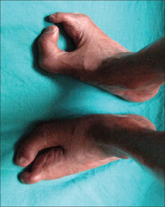
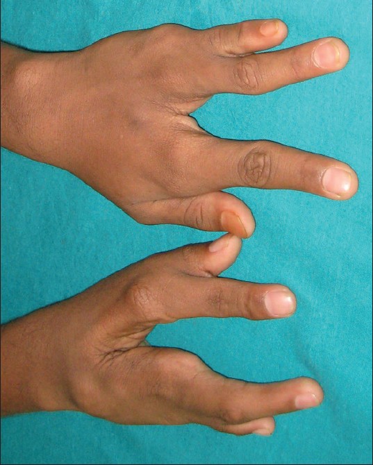
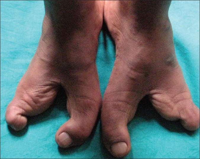
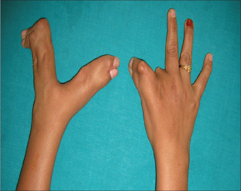
A 23-year-old female presented to the Outpatient Division, Department of Periodontics, Govt. Dental College, Kozhikode, Kerala, India [Figure 1] with congenital disorders on both limbs [Figure 2] and [Figure 3]. She was born as the third of six children of non-consanguineous parents after uncomplicated 37 weeks of gestation. Her 63-year old-father had congenital disorder of hands and feet [Figure 4] and [Figure 5]. Her 17-year-old younger brother also had congenital disorder of hands and feet [Figure 6] and [Figure 7], but her mother and other three siblings were normal. It was observed that the female subject , her father and younger brother had a genetic disorder termed as ectrodactyly ectodermal dysplasia- cleftting syndrome (EEC) syndrome. She was 155cm tall and weighed 47 kg and had nasal twang in her voice, coarse and sparse hair, sparse eyebrow, hypertelorism, syndactyly, ectrodactyly on both limbs, deep cleft on both limbs, and dysplastic nail of the left toe. Biochemical and hematological parameters, hormonal levels, blood hemoglobin level, and renal function were normal. Intelligence and other developmental milestones were also within the normal limits.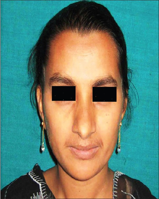 | Figure 1: Female patient with coarse and sparse hair, sparse eyebrow, and hypertelorism
Click here to view |
 | Figure 2: Hands with syndactyly, clinodactyly, and ectrodactyly of female patient
Click here to view |
 | Figure 3: Feet with syndactyly, clinodactyly, and ectrodactyly of female patient
Click here to view |
Oral examination
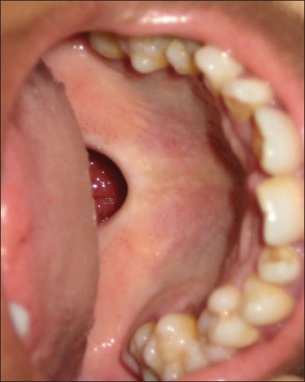
The patient had only 20 teeth [Figure 8] of which mandibular incisors had exfoliated a few months ago, and several other teeth were extracted due to mobility. She had soft palate cleft with missing uvula [Figure 9]. In both the jaws, teeth exhibited pathologic migration and mobility. Probing pocket depth measurements were performed on each tooth using a graduated Williams periodontal probe. Extreme probing depth measurements were noticed at maxillary right and left first molars and mandibular left first molar.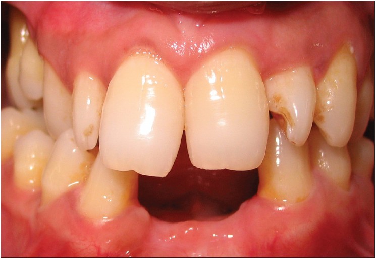 | Figure 8: Intraoral photograph of female patient demonstrating missing teeth
Click here to view |
 | Figure 9: Intraoral photograph of the patient Figure 10: OPG of the female patient demonstrating soft palate cleft with missing uvula of patient
Click here to view |
Panoramic radiograph [Figure 10] revealed generalized angular pattern of bone loss with greater loss around maxillary first molars, maxillary central incisors, maxillary second premolar, mandibular left second premolar, and first molar. Based on clinical and radiographic details, a diagnosis of generalized aggressive periodontitis was made.
The subject's father and brother were also diagnosed with generalized aggressive periodontitis.
| Discussion | | |
The ectodermal dysplasias are congenital heterogeneous group of inherited disorders with the primary defect in the development of embryonic ectodermal structures. Pure ectodermal dysplasias are manifested by defect in ectodermal structures alone, while ectodermal dysplasia "syndromes" like EEC syndrome have combination of ectodermal defects in association with other anomalies. Neuroectodermal and mesoectodermal derivatives and part of maxillofacial skeleton are also involved in EEC syndrome. This 23-year-old female patient manifested all the three classical features of EEC syndrome, which is a rare occurrence, while her family members (father and brother) showed variable expression of the EEC syndrome without the intraoral clefting. The etiology of EEC syndrome is genetic, and can be both familial and sporadic. Mutation in p63 gene [1] is found to be associated with EEC syndrome.
Several case reports of EEC syndrome have been published, [4],[5],[6] most of them pertaining to the dermatological, urogenital, and dental manifestations of EEC syndrome. The periodontal manifestations in EEC syndrome patients have never been focused in literature so far. The highly progressive and destructive nature of the bone loss around the teeth (aggressive periodontitis) might have developed at an early in life in these patients. Factors other than pathogenic microorganisms surrounding the tooth in the dental plaque could have influenced the actual clinical presentation. A genetic component might have also cumulated the pattern of periodontal disease in these patients. Quinn et al. in 1998 [9] reported that aggressive periodontitis may follow a dominant mode of transmission with the locus for aggressive periodontitis somewhere between the chromosomes number 3 to 7. On the other hand, autosomal recessive mode of transmission has also been noted for aggressive periodontitis. [10] Taken together, these studies indicate that both etiologic as well as genetic heterogenecity exist for aggressive periodontitis.
The gene for EEC syndrome is also found somewhere between chromosome 3 to 7. [3] There could be an overlapping association between locus on chromosomal group 3 to 7 for aggressive periodontitis and EEC syndrome, which could have accounted for the aggressive pattern of periodontal manifestations in these patients. This could open further avenues in investigating the overlapping association of genetics in EEC syndrome and aggressive periodontitis.
The 1999 classification of periodontal diseases and conditions have proposed various genetic disorders associated with periodontitis. Since no other reports exist in literature regarding the periodontal status of patients with EEC syndrome, EEC syndrome could be a new addition to the list of genetic disorders that predispose to periodontal tissue breakdown. It is essential that further research is needed to determine the genetic status of such patients. Management of cases with EEC syndrome and generalized aggressive periodontitis requires a multi-disciplinary approach.
| References | | |
| 1. | Celli J, Duijf P, Hamel BC, Bamshad M, Kramer B, Smits AP, et al. Heterozygous germline mutations in the p53 homolog p63 are the cause of EEC syndrome. Cell 1999;99:143-53. 
|
| 2. | Bixler D, Spivack J, Bennett J, Christian JC. The ectrodactyly-ectodermal dysplasia-clefting (EEC) syndrome.Report of 2 cases and review of the literature. Clin Genet 1972;3:43-51.
|
| 3. | Barrow LL, van Bokhoven H, Daack-Hirsch S, Andersen T,van Beersum SE, Gorlin R, et al. Analysis of the p63 gene in classical EEC syndrome, related syndromes, and non-syndromicorofacial clefts. J Med Genet 2002;39:559-66.
|
| 4. | Brunner HG, Hamel BC, BokhovenHv H. P63 gene mutations and human developmental syndromes. Am J Med Genet 2002;112:284-90.
|
| 5. | Peterson-Falzone SJ, Caldarelli DD, Landahl KL. Abnormal laryngeal vocal quality in ectodermal dysplasia. Arch Otolaryngol 1981;107:300-4.
|
| 6. | Ramirez D, Lammer EJ. Lacrimoauriculodentodigital syndrome with cleft lip/palate and renal manifestations. Cleft Palate Craniofac J 2004;41:501-6.
|
| 7. | Kinane DF. Periodontitis modified by systemic factors. Ann Periodontol 1999;4:54-64.
|
| 8. | Kinane DF, Hodge P, Eskdale J, Ellis R, Gallagher G. Analysis of genetic polymorphisms at the interleukin-10 and tumour necrosis factor loci in early-onset periodontitis. J Periodontal Res 1999;34:379-86.
|
| 9. | O'Quinn JR, Hennekam RC, Jorde LB, Bamshad M. Syndromicectrodactyly with severe limb, ectodermal, urogenital, and palatal defects maps to chromosome 19. Am J Hum Genet 1998;62:130-5.
|
| 10. | Marazita ML, Burmeister JA, Gunsolley JC, Koertge TE, Lake K, Schenkein HA. Evidence for autosomal dominant inheritance and race-specific heterogeneity in early-onset periodontitis. J Periodontol 1994;65:623-30.
|
[Figure 1], [Figure 2], [Figure 3], [Figure 4], [Figure 5], [Figure 6], [Figure 7], [Figure 8], [Figure 9], [Figure 10]
|