| |


 |
| Year : 2011 | Volume
: 5
| Issue : 3 | Page : 72-76 |
|
|
|
|
|
CASE REPORT Combined single photon emission computerized tomography and conventional computerized tomography: Clinical value for the shoulder surgeons?
Michael T Hirschmann, Rahel Schmid, Ranju Dhawan, Jiri Skarvan, Helmut Rasch, Niklaus F Friederich, Roger Emery
Department of Orthopaedic Surgery and Traumatology, Kantonsspital Bruderholz, Bruderholz, Switzerland
Correspondence Address:
Michael T Hirschmann
Department of Orthopaedic Surgery and Traumatology, Kantonsspital Bruderholz, Bruderholz
Switzerland
 Source of Support: None, Conflict of Interest: None  | 11 |
DOI: 10.4103/0973-6042.86242

|
|
|
|
| Date of Web Publication | 17-Oct-2011 |
 Abstract Abstract | | |
With the cases described, we strive to introduce single photon emission computerized tomography in combination with conventional computer tomography (SPECT/CT) to shoulder surgeons, illustrate the possible clinical value it may offer as new diagnostic radiologic modality, and discuss its limitations. SPECT/CT may facilitate the establishment of diagnosis, process of decision making, and further treatment for complex shoulder pathologies. Some of these advantages were highlighted in cases that are frequently seen in most shoulder clinics.
Keywords: Hemiarthroplasty, loosening, shoulder arthroplasty, SPECT/CT
How to cite this article:
Hirschmann MT, Schmid R, Dhawan R, Skarvan J, Rasch H, Friederich NF, Emery R. Combined single photon emission computerized tomography and conventional computerized tomography: Clinical value for the shoulder surgeons?. Int J Shoulder Surg 2011;5:72-6 |
How to cite this URL:
Hirschmann MT, Schmid R, Dhawan R, Skarvan J, Rasch H, Friederich NF, Emery R. Combined single photon emission computerized tomography and conventional computerized tomography: Clinical value for the shoulder surgeons?. Int J Shoulder Surg [serial online] 2011 [cited 2016 Apr 25];5:72-6. Available from: http://www.internationalshoulderjournal.org/text.asp?2011/5/3/72/86242 |
| Introduction | |  |
Single photon emission computerized tomography in combination with conventional computer tomography (SPECT/CT) pairs a high-resolution anatomical 3D-CT with a functional single photon emission tomography (SPECT). [1],[2],[3],[4],[5] Until recently, registration or image fusion of the functional (SPECT) with structural images (CT) has been difficult due to SPECT's poor spatial resolution (3-10 mm). Another important problem was the poor reproducibility in identifying the anatomical landmarks. [1],[6],[7] With the advent of integrated SPECT/CT machines in 1999, where the image data from SPECT are taken in the same coordinate frame as the CT scan, image fusion ceased to be an issue. Furthermore, hybrid SPECT/CT machines now provide superior SPECT data as the CT data can be used to derive the attenuation coefficients of the tissue. [8] With integrated machines, functional and anatomic information is provided in the same image, supporting diagnostic analysis of diseases where both types of data are vital. SPECT/CT has already proven useful as a diagnostic tool in areas such as endocrinology, cardiology, and neurosurgery. [1],[9],[10],[11],[12],[13],[14],[15],[16] In orthopedics, it has yet to be clinically validated and only a paucity of studies are available. [1],[2],[3],[4],[5],[17],[18],[19] With the cases described, we strive to introduce SPECT/CT to shoulder surgeons, illustrate the possible clinical value it may offer as new diagnostic radiologic modality, and discuss its limitations.
Technical description
Hybrid SPECT/CT was performed in two centers using either a hybrid system, Symbia T16 (Siemens, Erlangen, Germany) or Hawkeye-4 (GE Healthcare). The systems are equipped with a pair of low-energy, high-resolution collimators and incorporate a dual-head gamma camera with an integrated, 16- or 4- slice CT scanner. Patients were injected intravenously with 10-20 mCi (340-740 MBq) of 99m-technetium-labelled diphosphonate or 99m-technetium-labelled anti-granulocyte antibodies (CIS Bio International Sur Yvette, France). Perfusion images are obtained in the initial 60 seconds post-injection followed by blood pool images 2 to 5 minutes later. SPECT was performed at 3 to 5 hours, followed immediately by a CT with the patient in the same position.
| Case Reports | | |
Case 1
A 54-year-old male patient sustained a right-sided 4-part proximal humeral fracture, which was treated by open reduction and internal fixation with a Numelock polyaxial locking system (Stryker AG, Switzerland). One month later, he had to be revised due to a superficial wound infection with streptococcus. The infection resolved under antibiotic therapy with augmentin for 6 weeks. The plate was removed four months after initial fracture treatment.
At presentation to our clinic, the patient complained about activity-related pain and stiffness of his shoulder. Anterior-posterior and y-view radiographs revealed a valgus-impacted humeral head fragment. The healing of the fracture was in doubt and at least a partial humeral head necrosis was suspected [Figure 1]a. MRI showed wide intramedullary osteolytic zones, which were attributed either to a pseudoarthrosis or recurrence of infection. In addition, a severe osteoarthritis and a fatty degeneration of the supraspinatus muscle (Goutallier 2) were identified [Figure 1]b. To exclude a persistence of an infection, an anti-granulocyte SPECT/CT was performed. This showed absent tracer uptake of the humeral head fragment which was attributed to a humeral head necrosis. No signs of infection were present [Figure 1]c. The patient was scheduled for total shoulder arthroplasty.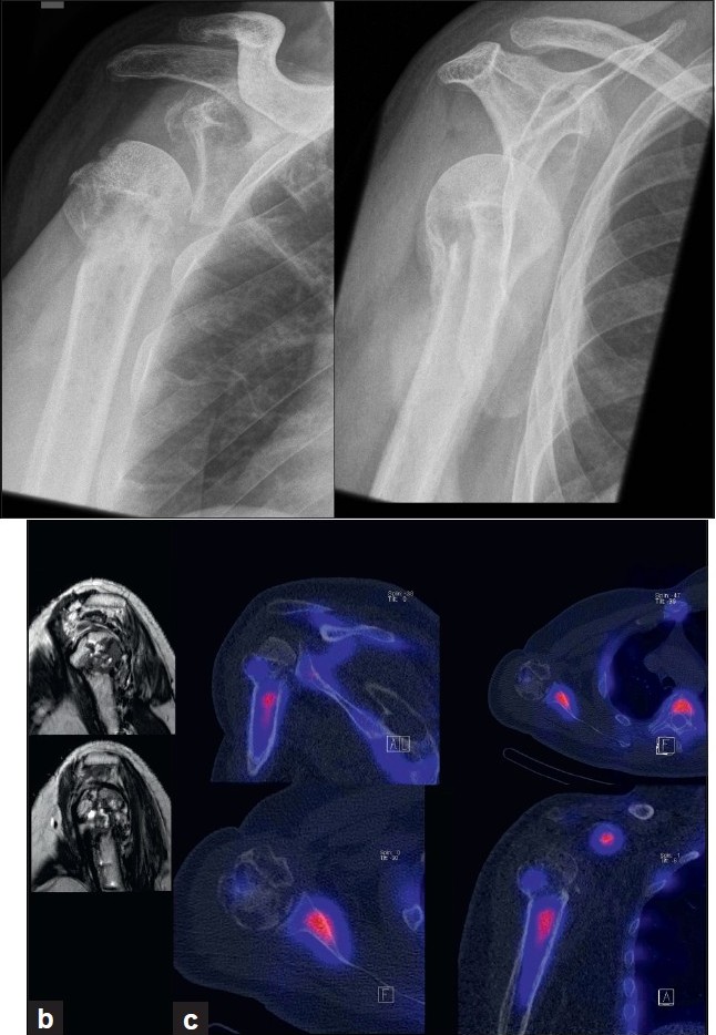 | Figure 1: (a)Anterior-posterior and y-view radiographs of the right shoulder at initial presentation indicating a valgus-impacted humeral head fragment and doubtful partial osteonecrosis of the humeral head
Figure 1: (b) MRI showing extensive cystic changes within the humeral head; (c) 99mTc-HDP-SPECT/CT showing decreased uptake of the humeral head fragment indicating a humeral head necrosis
Click here to view |
Case 2
A 70-year-old female patient underwent an inverse shoulder arthroplasty (ANATOMICAL 3, Zimmer, Switzerland) due to a cuff arthropathy. Seven months later, she suffered from activity-related pain in her shoulder. Anterior posterior and y-view radiographs showed extensive osteolysis around the glenoid component [Figure 2]a. In SPECT/CT, increased tracer uptake between the glenoid component and the host bone was present [Figure 2]b. There was no increased tracer uptake around the stem indicating a well-fixed humeral stem component. At revision surgery, the glenoid component was loose. It was revised to a bipolar humeral head (custom-made, Zimmer, Switzerland) and the host bone defect was filled with autologous cancellous bone graft from the ipsilateral iliac crest. The stem was not revised, as it was well fixed [Figure 2]c. At three-month follow-up, the patient had not regained full range of motion, but presented with less in activities of daily living.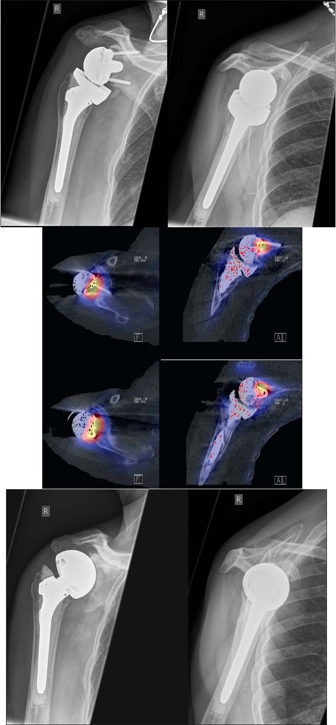 | Figure 2: (a)Anterior-posterior and y-view radiographs of the right shoulder showing wide radiolucent lines around the glenoid peg
Figure 2b: 99mTc-HDP-SPECT/CT showing increased tracer uptake at the glenoid component-host bone interface, which was interpreted as clear sign for mechanical loosening
Figure 2c: Anterior-posterior and y-view radiographs of the right shoulder after revision to a bipolar cup prosthesis
Click here to view |
Case 3
A 59-year-old male patient underwent a shoulder hemiarthroplasty (EPOCA, Synthes, Switzerland) due to a 4-part proximal humeral fracture. The postoperative recovery was uneventful until 3 years postoperatively when shoulder pain increased. Anterior-posterior and y-view radiographs showed osteolytic zones around the humeral stem indicating aseptic loosening of the hemiarthroplasty [Figure 3]a. Since osteolytic zones and clinical symptoms often poorly correlate in shoulder arthroplasty, a SPECT/CT scan was performed. It showed increased tracer uptake only around the stem in all three phases, which was attributed to mechanical loosening [Figure 3]b. The hemiarthroplasty was revised (GLOBAL, DePuy, Switzerland) and a satisfying recovery was obtained.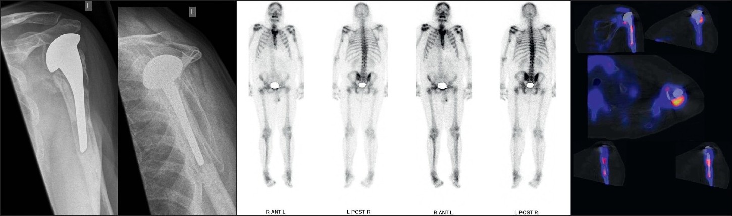 | Figure 3: (a)Anterior-posterior and y-view radiographs of the left shoulder indicating radiolucent lines around the humeral stem
Figure 3b: Planar scintigraphy and 99mTc-HDP-SPECT/CT showing increased tracer uptake around the humeral stem, which was caused by mechanical loosening
Click here to view |
Case 4
A 58-year-old patient presented with a painful right shoulder with reduced range of movement and a history of arthroscopy 3 months previously for a stiff painful glenohumeral joint. Given history of the arthroscopy and hence concern for infection, a two-phase bone scan supplemented with SPECT-CT (GE, Hawkeye-4) was performed. Accentuated blood pool activity was found on delayed planar scans with tracer uptake at the right humeral head. Although this finding supported osteonecrosis, the SPECT-CT [Figure 4] localized the activity more specifically at the glenohumeral joint interface. In addition, the CT showed radiographic erosive changes at both subarticular joint surfaces confirming a primary joint-based process [Figure 4]. A total shoulder replacement (TSR) was performed. No bacterial growth was confirmed in culture specimens. After a symptom-free period of 9 months, pain recurred. On examination, the pain was thought to be of subacromial origin and ultrasound-guided injections of the subacromial bursa brought partial relief. Continuing pain prompted the surgeon to consider prosthetic loosening. A CT scan in a modified position [20] showed no adverse features [Figure 4]. A SPECT/CT at 14 months after TSR showed no significant early blood pool phase activity mitigating against infection, whilst delayed planar images showed mild periprosthetic activity considered to be in the realm of osseous remodeling after TSR. More focal tracer activity was present at the acromion, raising this site as possible pain generators [Figure 4]. An arthroscopic subacromial decompression resulted in significant pain relief as assessed on clinical follow-up at one-month post-procedure.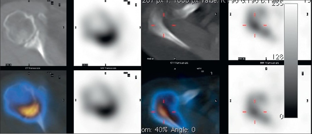 | Figure 4: (a)SPECT/CT showing erosive arthropathy with joint-based tracer uptake
Figure 4b: CT showing no significant periprosthetic lucency; planar scintigraphy indicated normal blood pool activity, delayed moderate periprosthetic tracer activity;SPECT/CT clearly found increased tracer activity of the acromion and acromioclavicular joint
Click here to view |
| Discussion | | |
The most important finding of the present case study was that SPECT/CT may facilitate the establishment of diagnosis, process of decision making, and further treatment for complex shoulder pathologies. Some of these advantages were highlighted in cases that are frequently seen in most shoulder clinics.
In case one, the anti-granulocyte SPECT/CT helped to noninvasively distinguish recurrence of infection from humeral head necrosis. 99m-Tc-labeled anti-granulocyte antibodies are directed against the concomitant infiltration of granulocytes in infected tissues and thereby these are helpful in diagnosing and localizing joint infections. [13],[21] Recently, Graute et al. assessed the diagnostic sensitivity of scintigraphy, SPECT, and SPECT/CT. In relation to planar scintigraphy, SPECT/CT substantially improved the sensitivity and specificity for diagnosis and localization of suspected joint infections. [21] Although the authors only assessed infections of knee and hip arthroplasty, it could be speculated that similar results might be found in patients after shoulder arthroplasty.
The second case illustrates a common problem after shoulder arthroplasty, namely glenoid loosening. In patients after reversed shoulder prosthesis as well as after conventional arthroplasty, glenoid loosening represents one of the most common failure modes causing postoperative pain, limited function, and the need for revision surgery. [20] To date, detection and monitoring of glenoid fixation failure is considered to be difficult and shows a great inter- and intraobserver variability. [22] Radiolucent lines, particularly the width, are difficult to interpret in conventional radiographs. This might be due to inaccurate positioning of the patient and anatomical variation in glenoid version and mobility of the shoulder girdle. [22] Clearly, the radiolucent lines appeared to be smaller when retroversion was 10 or more and when anteversion was 15 or more. [22]
Nowadays, three-dimensional reconstructions of CT provide the surgeon with a better 3D understanding of patient's anatomy. In addition, the position and orientation of the prosthetic components after shoulder arthroplasty could be assessed in relation to standardized frames of reference. With SPECT/CT, it is not only possible to determine the width of radiolucencies and the localization of the metabolic tracer uptake, but also assess the orientation of the shoulder prosthesis, in particular the glenoid component. The orientation of the glenoid component has been reported to be a risk factor for early glenoid loosening. [23]
The potential benefit that SPECT/CT might provide for patients after shoulder arthroplasty is further highlighted by the third case where SPECT/CT identified osteolysis around the humeral stem and metabolic tracer uptake reflecting toggling of the component as cause of the patient's pain. SPECT-CT may be especially useful when MRI is contraindicated, equivocal, or prone to artefacts when metalwork is in situ, such as in patients after arthroplasty.
The fourth case highlights the potential clinical benefit that SPECT/CT might offer in patients with shoulder pain. As the shoulder girdle consists of several joints and different anatomical structures within, a small area pain can very often not be localized to distinct structures or joints. SPECT/CT might help to identify the pain generator, which was shown in the presented case.
Limitations
Using SPECT/CT in orthopedic patients, one should be aware of its limitations. As this was only a small case series, conclusions should be drawn with all due caution. To date, no sufficient information about the natural course of SPECT/CT after shoulder surgery has been established. The sensitivity and specificity of SPECT/CT for establishing the diagnosis in patients with shoulder problems has not been investigated to date as there is only a paucity of studies available in orthopedics. [2],[3],[4],[17],[18],[19] The cost-effectiveness of SPECT/CT compared with other imaging has not been addressed so far.
| Conclusions | | |
SPECT/CT provides shoulder surgeons with a promising diagnostic tool, which may be particularly helpful in patients with shoulder pain, in which radiological diagnostic possibilities are currently limited, such as painful partial or total shoulder arthroplasty. The clinical value, the sensitivity and specificity of SPECT/CT in patients before and after shoulder surgery should be further investigated.
| References | | |
| 1. | Bybel B, Brunken RC, DiFilippo FP, Neumann DR, Wu G, Cerqueira MD. SPECT/CT imaging: Clinical utility of an emerging technology. Radiographics 2008;28:1097-113. 
|
| 2. | Hirschmann MT, Iranpour F, Davda K, Rasch H, Huegli R, Friederich NF. Combined single-photon emission computerized tomography and conventional computerized tomography (SPECT/CT): Clinical for the knee surgeons? Knee Surg Sports Traumatol Arthrosc 2010;18:341-5.
|
| 3. | Hirschmann MT, Iranpour F, Konala P, Kerner A, Rasch H, Cobb JP, et al. A novel standardized algorithm for evaluating patients with painful total knee arthroplasty using combined single photon emission tomography and conventional computerized tomography. Knee Surg Sports Traumatol Arthrosc 2010;18:939-44.
|
| 4. | Mohan HK, Gnanasegaran G, Vijayanathan S, Fogelman I. SPECT/CT in imaging foot and ankle pathology- the demise of other coregistration techniques. Semin Nucl Med 2010;40:41-51.
|
| 5. | Scharf S. SPECT/CT imaging in general orthopaedic practice. Semin Nucl Med 2009;39:293-307.
|
| 6. | Ahmad R, Kumar GS, Katam K, Dunlop D, Pozo JL. Significance of a "hot patella" in total knee replacement without primary patellar resurfacing. Knee 2009;16:337-40.
|
| 7. | Schillaci O. Hybrid SPECT/CT: A new era for SPECT imaging? Eur J Nucl Med Mol Imaging 2005;32:521-4.
|
| 8. | Patton JA, Turkington TG. SPECT/CT physical principles and attenuation correction. J Nucl Med Technol 2008;36:1-10.
|
| 9. | Bailey E, HoShon I, Roach P. Additional benefit of SPECT/CT in bone scanning of metastatic renal cell carcinoma. Clin Nucl Med 2007;32:411-4.
|
| 10. | Bockisch A, Freudenberg LS, Schmidt D, Kuwert T. Hybrid imaging by SPECT/CT and PET/CT: Proven outcomes in cancer imaging. Semin Nucl Med 2009;39:276-89.
|
| 11. | Chowdhury FU, Scarsbrook AF. The role of hybrid SPECT/CT in oncology: Current and emerging clinical applications. Clin Radiol 2008;63:241-51.
|
| 12. | D`Amico A, Szczucka K, Borys D, Gorczewski K, Steinhof K. SPECT/CT fusion: A new diagnostic tool for endocrinology. Endokrynol Pol 2006;57:4.
|
| 13. | Filippi L, Schillaci O. Usefulness of hybrid SPECT/CT in 99mTc-HMPAO-labeled leukocyte scintigraphy for bone and joint infections. J Nucl Med 2006;47:1908-13.
|
| 14. | Filippi L, Uccioli L, Giurato L, Schillaci O. Diabetic foot infection: Usefulness of SPECT/CT for 99m-Tc-HMPAO-labeled leukocyte imaging. J Nucl Med 2009;50:1042-6.
|
| 15. | Mahmarian JJ. Hybrid SPECT/CT: Integration of CT coronary artery calcium scoring and angiography with myocardial perfusion. Curr Cardiol Rep 2007;9:129-35.
|
| 16. | Van der Ploeg IM, Valdes Olmos RA, Kroon BB, Nieweg OE. The hybrid SPECT/CT as an additional lymphatic mapping tool in patients with breast cancer. World J Surg 2008;32:1930-4.
|
| 17. | Knupp M, Pagenstert GI, Barg A, Bolliger L, Easley ME, Hintermann B. SPECT/CT compared with conventional imaging modalities for the assessment of the carus and valgus malaligned hindfoot. J Orthop Res 2009;27:1461-6.
|
| 18. | Hirschmann MT, Konala P, Iranpour F, Kerner A, Rasch H, Friederich NF. Clinical value of SPECT/CT for evaluation of patients with painful knees after total knee arthroplasty - a new dimension of diagnostics? BMC Musculoskelet Disord 2011;12:36.
|
| 19. | Papathanassiou D, Bruna-Muraille C, Jouannaud C, Gagneux-Lemoussu K, Eschard JP, Liehn JC. Single-photon emission computed tomography combined with computed tomography (SPECT/CT) in bone diseases. Joint Bone Spine 2009;76:474-80.
|
| 20. | Gregory T, Hansen U, Taillieu F, Baring T, Brassart N, Mutchler C, et al. Glenoid loosening after total shoulder arthroplasty: An in vitro CT-scan study. J Orthop Res 2009;27:1589-95.
|
| 21. | Graute V, Feist M, Lehner S, Haug A, Müller PE, Bartenstein P, et al. Detection of low-grade prosthetic joint infections using (99m)Tc-antigranulocyte SPECT/CT: Initial clinical results. Eur J Nucl Med Mol Imaging 2010;37:1751-9.
|
| 22. | Havig MT, Kumar A, Carpenter W, Seiler JG 3 rd . Assessment of radiolucent lines about the glenoid. An in vitro radiographic study. J Bone Joint Surg Am 1997;79:428-32.
|
| 23. | Farron A, Terrier A, Buchler P. Risks of loosening of a prosthetic glenoid implanted in retroversion. J Shoulder Elbow Surg 2006;15:521-6.
|
[Figure 1], [Figure 2], [Figure 3], [Figure 4]
| This article has been cited by | | 1 |
Effect of high tibial osteotomy on joint loading in symptomatic patients with varus aligned knees: a study using SPECT/CT |
|
| Armin Mucha,Milos Dordevic,Anna Hirschmann,Helmut Rasch,Felix Amsler,Markus P. Arnold,Michael T. Hirschmann | | Knee Surgery, Sports Traumatology, Arthroscopy. 2014; | | [Pubmed] | [DOI] | | | 2 |
SPECT-CT: applications in musculoskeletal radiology |
|
| S Saha,C Burke,A Desai,S Vijayanathan,G Gnanasegaran | | The British Journal of Radiology. 2013; 86(1031): 20120519 | | [Pubmed] | [DOI] | | | 3 |
4D-SPECT/CT in orthopaedics: a new method of combined quantitative volumetric 3D analysis of SPECT/CT tracer uptake and component position measurements in patients after total knee arthroplasty |
|
| Helmut Rasch,Anna L. Falkowski,Flavio Forrer,Johann Henckel,Michael T. Hirschmann | | Skeletal Radiology. 2013; 42(9): 1215 | | [Pubmed] | [DOI] | | | 4 |
Assessment of loading history of compartments in the knee using bone SPECT/CT: A study combining alignment and 99mTc-HDP tracer uptake/distribution patterns |
|
| Hirschmann, M.T. and Schön, S. and Afifi, F.K. and Amsler, F. and Rasch, H. and Friederich, N.F. and Arnold, M.P. | | Journal of Orthopaedic Research. 2013; 31(2): 268-274 | | [Pubmed] | | | 5 |
Clinical applications of SPECT/CT in imaging the extremities |
|
| Martin W. Huellner,Klaus Strobel | | European Journal of Nuclear Medicine and Molecular Imaging. 2013; | | [Pubmed] | [DOI] | | | 6 |
Radiographically Occult and Subtle Fractures: A Pictorial Review |
|
| Mohamed Jarraya,Daichi Hayashi,Frank W. Roemer,Michel D. Crema,Luis Diaz,Jane Conlin,Monica D. Marra,Nabil Jomaah,Ali Guermazi | | Radiology Research and Practice. 2013; 2013: 1 | | [Pubmed] | [DOI] | | | 7 |
Assessment of loading history of compartments in the knee using bone SPECT/CT: A study combining alignment and 99mTc-HDP tracer uptake/distribution patterns |
|
| Michael T. Hirschmann,Stephan Schön,Faik K. Afifi,Felix Amsler,Helmut Rasch,Niklaus F. Friederich,Markus P. Arnold | | Journal of Orthopaedic Research. 2013; 31(2): 268 | | [Pubmed] | [DOI] | | | 8 |
SPECT/CT in patients with painful knee arthroplasty—what is the evidence? |
|
| Michael T. Hirschmann,Johann Henckel,Helmut Rasch | | Skeletal Radiology. 2013; 42(9): 1201 | | [Pubmed] | [DOI] | | | 9 |
SPECT/CT in der Handgelenkdiagnostik |
|
| M.W. Huellner,K. Strobel,U. Hug,U. Wartburg,P. Veit-Haibach | | Der Radiologe. 2012; 52(7): 621 | | [Pubmed] | [DOI] | | | 10 |
Diagnostic and therapeutic impact of SPECT/CT in patients with unspecific pain of the hand and wrist |
|
| Schleich, F.S. and Schürch, M. and Huellner, M.W. and Hug, U. and von Wartburg, U. and Strobel, K. and Veit-Haibach, P. | | EJNMMI Research. 2012; 2(1): 1-8 | | [Pubmed] | | | 11 |
SPECT/CT in diagnostics of the hand joint [SPECT/CT in der handgelenkdiagnostik] |
|
| Huellner, M.W. and Strobel, K. and Hug, U. and Von Wartburg, U. and Veit-Haibach, P. | | Radiologe. 2012; 52(7): 621-628 | | [Pubmed] | | | 12 |
Diagnostic and therapeutic impact of SPECT/CT in patients with unspecific pain of the hand and wrist |
|
| Florian S Schleich,Maja Schürch,Martin W Huellner,Urs Hug,Urs von Wartburg,Klaus Strobel,Patrick Veit-Haibach | | EJNMMI Research. 2012; 2(1): 53 | | [Pubmed] | [DOI] | |
|
|
|


|
|
|
|
|


