| |


 |
| Year : 2013 | Volume
: 7
| Issue : 2 | Page : 79-82 |
|
|
|
|
|
SURGICAL TECHNIQUE Arthroscopic fixation with a minimally invasive axillary approach for latissimus dorsi transfer using an endobutton in massive and irreparable postero-superior cuff tears
Yariv Goldstein1, Jean Grimberg2, Philippe Valenti3, Ofir Chechik4, Michael Drexler4, Jean Kany5
1 Department of Orthopedic Surgery, The Shoulder Unit, Tel Aviv Sourasky Medical Center, Tel Aviv, Israel; Department of Upper Extremity, Clinique De l'Union, Toulouse, France,
2 Department of Orthopedic Surgery, Clinique des Lilas, Paris, France,
3 Clinique Jouvenet, Institut de la main, Paris, France,
4 Department of Orthopedic Surgery, The Shoulder Unit, Tel Aviv Sourasky Medical Center, Tel Aviv, Israel,
5 Department of Upper Extremity, Clinique De l'Union, Toulouse, France,
Correspondence Address:
Yariv Goldstein
Boulevard de Ratalens, 31240, Saint Jean, France
 Source of Support: None, Conflict of Interest: None  | 6 |
DOI: 10.4103/0973-6042.114223

|
|
|
|
| Date of Web Publication | 29-Jun-2013 |
 Abstract Abstract | | |
Arthroscopically assisted latissimus dorsi transfer is a viable option for treatment of patients in their 50s to 70s, without arthritis of the glenohumeral joint, who suffer from massive rotator cuff tears that are not amendable to primary repair due to fatty changes in the muscle tissue, or that have failed previous repair attempts. This procedure offers immediate and dramatic pain relief and is not as technically demanding as one might think. Understanding and respecting the principles of tendon transfer is a key to the success of this procedure.
Keywords: Latissimus dorsi, massive cuff tear endobutton, shoulder arthroscopy, tendon transfer
How to cite this article:
Goldstein Y, Grimberg J, Valenti P, Chechik O, Drexler M, Kany J. Arthroscopic fixation with a minimally invasive axillary approach for latissimus dorsi transfer using an endobutton in massive and irreparable postero-superior cuff tears. Int J Shoulder Surg 2013;7:79-82 |
How to cite this URL:
Goldstein Y, Grimberg J, Valenti P, Chechik O, Drexler M, Kany J. Arthroscopic fixation with a minimally invasive axillary approach for latissimus dorsi transfer using an endobutton in massive and irreparable postero-superior cuff tears. Int J Shoulder Surg [serial online] 2013 [cited 2016 May 22];7:79-82. Available from: http://www.internationalshoulderjournal.org/text.asp?2013/7/2/79/114223 |
| Introduction | |  |
This article describes the evolution of our previously reported technique for the arthroscopically assisted latissimus dorsi (LD) Transfer and fixation with an interference screw. [1] During the years, transfer of the LD was used as a salvage procedure to massive irreparable rotator cuff tears. The technique was first described by Christian Gerber in his 1988 article. [2] Since then many alterations to the original technique were used, mainly altering the anchorage technique with the use of suture anchors, bone tunnels, Interference screws or a combination of techniques. The LD was regarded as a large vascularized tendon that can be used to close a massive cuff defect and that exerts an external rotation and head depressing moment. [2],[3],[4],[5],[6] The transfer of the LD tendon, to our knowledge, was never regarded as a tendon transfer per-se and therefore, less notice was given to the principles of tendon transfer. This could account for inconsistent outcomes.
Five important principles of tendon transfer are:
- The point of fixation - precise location of the point of fixation of the transferred tendon
- The muscle tension - the tension acquired at the end of the operation should resemble the original tension within the transferred muscle
- Tendon to bone contact - fixation to the bone at the site of insertion should provide as much contact as possible between the transferred tendon and the surrounding bone
- Minimal exposure and tissue damage
- The use of a muscle that acts as an agonist muscle to the one it is replacing.
In our previous publication, we described an arthroscopically assisted LD transfer. [1] For fixation, we used either titanium or absorbable interference screws (IFS) (Stryker Interference Screw, www.stryker.com). Using the IFS technique has two shortcomings:
First, we had to drill the tunnel for the tendon in a cranial to caudal direction. This puts the acromion in the way, especially, when the patient has a large lateral acromion extension, as described by Nyffeler et al. [7] In these cases, even the use of the neviaser portal does not offer a clear path to the humeral head at the intended insertion point. Second, as the IFS has a diameter of 7-8 mm, mal-positioning of the drill hole laterally resulted in a stress riser that led to the fracture of the lateral cortex and failure of fixation in two cases. These patients required revision surgery in which the tendon was re-attached using suture anchors.
Drilling the tunnel in a caudal to cranial direction addressed the first issue as the acromion was now out of the way. We still had to deal with the diameter of the tunnel required for our fixation device.
In our exploration for a safer way to anchor the transferred tendon to the Humerus, we thought of using an endobutton against the anterolateral cortex of the Humerus, on the lateral aspect of the bicipital groove.
This technique has three obvious advantages over former techniques:
- The tunnel drilled through the Humerus should match the tubularized tendon diameter. This ensures that the transferred tendon is in full contact with the surrounding bone tunnel
- Using an endobutton allows us to precisely adjust the tension of the tendon prior to fixation
- A metallic button laid against the anterolateral cortex of the Humerus is a strong fixation technique relying on cortical bone which is inherently stronger than cancellous bone.
| Surgical Technique | | |
The patient is placed in the standard lateral decubitus position with 3 kg of traction applied to his arm using weights and a pulley system. A 5 cm incision (4-7 depending on patient anatomy) is made at the anterior (axillary) border of the scapula, 5 cm cranial to the scapular apex [Figure 1]. The incision can be easily extended caudally or cranially as needed.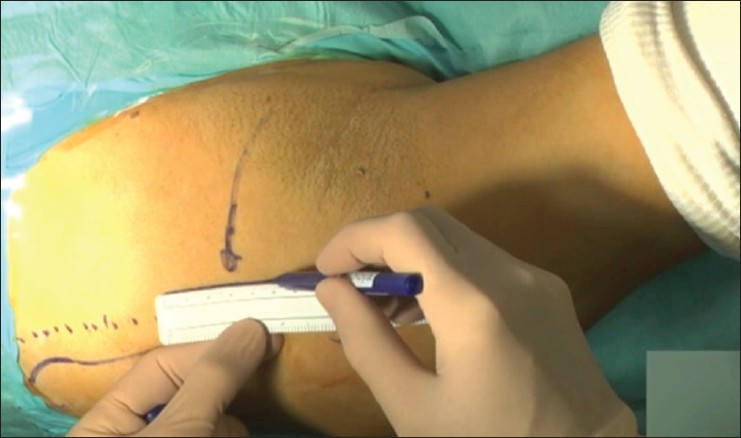 | Figure 1: A 5 cm incision (4-7 depending on patient anatomy) is made at the anterior (axillary) border of the scapula, 5 cm cranial to the scapular apex
Click here to view |
Once the incision is made, the first visible muscle is the LD. The redundancy of skin around the axilla is used to extend the dissection subcutaneously.
The next step is the identification and mobilization of the neurovascular pedicle of the LD, which enters the muscle mid belly from its medial surface [Figure 2]. Thorough mobilization is required to prevent over stretching this structure while the transfer is performed and tension is placed over the muscle-tendon flap. A healed but clinically and electrically non-functional transfer, as described by Codsi et al. [8] can be a result of insufficient release around the pedicle of the LD. | Figure 2: Identification and mobilization of the neurovascular pedicle of the latissimus dorsi which enters the muscle mid belly from its medial surface
Click here to view |
Once the pedicle is mobile, the LD muscle belly is separated from the muscle belly of the Teres major (TM). Beck and Hoffer performed a cadaveric study and described the tendons of the LD and the TM as a conjoined tendon. [9] Goldberg found fascial connections between the muscle bellies of the muscles in all the specimens of his cadaveric study. [10] In our experience, a fatty interface is present in about 50% of the cases between the two muscles and can be used for separating the fascial connections. Care should be taken to thoroughly release these fascial connections between the LD muscle belly and that of the TM. Once the pedicle has been released and the muscle freed from its surrounding structures, the tendon should be carefully followed distally to its humeral insertion. Effort should be made to achieve a graft that is as long as possible. The release of the tendon should be done at the tendon-bone transition zone. This allows for up to 2 cm of additional tendon tissue. Prior to sectioning of the tendon, we place two marker sutures on the muscle belly at a distance of 3 cm from one another while the arm is at an abducted and externally rotated position. This position puts the LD muscle at maximum tension. These marker sutures are used later on to restore tension to the muscle belly when the tendon is being fixed. Restoring the correct tension of the muscle belly is important for muscle activity but we believe that restoring the correct tension on the muscle-tendon unit and on the neurovascular pedicle is crucial in maintaining a viable functional flap.
The tenotomy is performed in a cranial-caudal manner. Due to the close proximity of the deep axillary vessels medially, this should be done with extreme care.
The tendon of the LD is a flat thin structure. When used to close a massive cuff defect this is advantageous. Our technique requires a structure that is tubular. We therefore tubularize the tendon using two sutures (Arthrex Fiberloop No. 2, Arthrex Inc., FL USA, Tornier Force Fiber, Tornier Inc.), total of four suture limbs.
We normally achieve a tubular tendon measuring about 7 mm in diameter and 7 cm in length [Figure 3]. This is consistent with the publication by Goldberg et al. [10] | Figure 3: We normally achieve a tubular tendon measuring about 7 mm in diameter and 7 cm in length
Click here to view |
At this stage, we further release the muscle belly bluntly, all the way to the apex of the scapula while maintaining traction on the now tabularized tendon. The goal of the release is reached when one's finger could easily engulf the muscle belly circumferentially.
The next step comprises the preparation of the route for the transferred tendon. Creating a tunnel that allows for the shortest route for the transfer is preferable because it preserves the muscle-tendon length.
This step begins with an arthroscopic preparation of the greater tuberosity via the standard portals (posterior, anterolateral and lateral portal for visualization and instrumentation). Next, the scope is moved in a posterior direction over the Teres minor in a caudal direction. The plane between the Teres minor, when intact, and the deltoid is developed using elctrocoagulation and an alternating shaver blade. Once possible, the suture manipulator is passed through this newly created space and retrieved through the axillary incision. The four suture limbs of the tabularized tendon are retrieved and passed through the tunnel and the tendon is carefully pulled while the index finger helps in creating the passage. The diameter of the tubular tendon is measured using a standard metallic diameter gauge.
The next step is the drilling of the bone tunnel. We introduce a specially designed two parts guide, where the proximal part is placed over the humeral head and guided to the desired position of insertion under vision using the scope. The distal part is then placed against the cortex of the humerus at the desired exit point of the tunnel. The two parts of the guide are then attached and secured. This device allows us to precisely choose our entry and exit points and to hold the position while drilling and passing the shuttle guide [Figure 4]. | Figure 4: Specially designed guide aids positioning of the insertion point and allows for retrograde drilling of the tunnel
Click here to view |
A shuttle guide is then placed in a caudal to cranial direction. A drill with the same diameter as the tendon graft is introduced over the shuttle guide and drilling is performed in the same direction (caudal to cranial). The sutures are then passed with the aid of the shuttle guide from cranial to caudal and retrieved outside the skin, where the button is attached [Figure 5]. While maintaining the precise tension of the muscle-tendon unit using the marker sutures placed before sectioning of the tendon, the button is lowered gradually until it lays flush against the bone cortex. The sutures are then tied with the aid of a knot pusher. Dynamic function of the graft is assessed with great care. | Figure 5: Endobutton is introduced through the incision used for positioning of the guide. Tension is maintained while the device is fixed
Click here to view |
| Discussion | | |
With the advancement in treatment options for repairable rotator cuff RC tears and for (RC) arthropathy, we still face a problem when we look for treatment for an ever growing group of patients in their 50s to 70s, without arthritis of the glenohumeral joint, who suffer from massive rotator cuff tears that are not amendable to primary repair due to fatty changes in the muscle tissue, or that have failed previous repair attempts. These people pursue an active life style and many times, have good passive (and sometimes active) range of motion, but are in constant pain.
We believe that a transfer of the LD has much to offer to this specific group of patients. We perform this surgery with good results on patients that have not yet been operated on as well as on patients who had failed previous attempts of rotator cuff repair. We manage to respect four out of the five principles of tendon transfer.
Our Arthroscopic technique is much less invasive than the classic LD transfer [2] or the single incision technique. [4] It is relatively easy to perform and in our experience offers immediate and dramatic pain relief.
The use of an arthroscope and a specially designed guide allows us to accurately choose our insertion point into the humeral head, with minimal exposure and damage to surrounding tissue [Figure 4]. The tunnel is drilled at the same diameter of the tabularized tendon, which allows for a large surface area of contact between the bone and the tendon. This increases the chance for good healing of tendon into the insertion point.
The use of an endobutton as a fixation device allows us to have more precise control over the tension of the transferred muscle-tendon unit. Once the desired tension is recreated and maintained, the button is locked against the lateral cortex of the bicipital groove, a strong cortical structure [Figure 5].
Being an antagonist muscle to external rotation, the LD is not an agonist muscle and the 5 th principle is therefore, not respected.
Our experience is showing promise after 1 year but longer follow-up is needed as we document improvement in results as late as 3 years after surgery.
| References | | |
| 1. | Kany J, Kumar HA, Chang V, Grimberg J, Garret J, Valenti P. Mini invasive axillary approach and arthroscopic humeral head interference screw fixation for latissimus dorsi transfer in massive and irreparable posterosuperior rotator cuff tears. Techn ShElbow Surg 2010;11:8-14. 
|
| 2. | Gerber C, Vinh TS, Hertel R, Hess CW. Latissimus dorsi transfer for the treatment of massive tears of the rotator cuff. A preliminary report. Clin Orthop Relat Res 1988;232:51-61.
[PUBMED] |
| 3. | Habermeyer P, Magosch P, Rudolph T, Lichtenberg S, Liem D. Transfer of the tendon of latissimus dorsi for the treatment of massive tears of the rotator cuff: A new single-incision technique. J Bone Joint Surg Br 2006;88:208-12.
[PUBMED] |
| 4. | Lehmann LJ, Mauerman E, Strube T, Laibacher K, Scharf HP. Modified minimally invasive latissimus dorsi transfer in the treatment of massive rotator cuff tears: A two-year follow-up of 26 consecutive patients. Int Orthop 2010;34:377-83.
[PUBMED] |
| 5. | Nové-Josserand L, Costa P, Liotard JP, Safar JF, Walch G, Zilber S. Results of latissimus dorsi tendon transfer for irreparable cuff tears. Orthop Traumatol Surg Res 2009;95:108-13.
|
| 6. | Moursy M, Forstner R, Koller H, Resch H, Tauber M. Latissimus dorsi tendon transfer for irreparable rotator cuff tears: A modified technique to improve tendon transfer integrity. J Bone Joint Surg Am 2009;91:1924-31.
[PUBMED] |
| 7. | Nyffeler RW, Werner CM, Sukthankar A, Schmid MR, Gerber C. Association of a large lateral extension of the acromion with rotator cuff tears. J Bone Joint Surg Am 2006;88:800-5.
[PUBMED] |
| 8. | Codsi MJ, Hennigan S, Herzog R, Kella S, Kelley M, Leggin B, et al. Latissimus dorsi tendon transfer for irreparable posterosuperior rotator cuff tears. Surgical technique. J Bone Joint Surg Am 2007;89 suppl 2 Pt. 1:1-9.
|
| 9. | Beck PA, Hoffer MM. Latissimus dorsi and teres major tendons: Separate or conjoint tendons? J Pediatr Orthop 1989;9:308-9.
[PUBMED] |
| 10. | Goldberg BA, Elhassan B, Marciniak S, Dunn JH. Surgical anatomy of latissimus dorsi muscle in transfers about the shoulder. Am J Orthop (Belle Mead NJ) 2009;38:E64-7.
[PUBMED] |
[Figure 1], [Figure 2], [Figure 3], [Figure 4], [Figure 5]
| This article has been cited by | | 1 |
Latissimus dorsi tendon transfer for irreparable postero-superior cuff tears: current concepts, indications, and recent advances |
|
| Jean Grimberg,Jean Kany | | Current Reviews in Musculoskeletal Medicine. 2014; | | [Pubmed] | [DOI] | | | 2 |
Clinic and electromyographic results of latissimus dorsi transfer for irreparable posterosuperior rotator cuff tears |
|
| Ricardo De Casas,Matías Lois,Myriam Cidoncha,Miguel Valadron | | Journal of Orthopaedic Surgery and Research. 2014; 9(1) | | [Pubmed] | [DOI] | |
|
|
|
|


